

Ectopic pancreatic-type malignancy presenting in a Meckel's diverticulum: a case report and review of the literature
- PMID: 19545406
- PMCID: PMC2709896
- DOI: 10.1186/1477-7819-7-54
Ectopic pancreatic-type malignancy presenting in a Meckel's diverticulum: a case report and review of the literature
Abstract
Background: Neoplasms arising from Meckel's diverticulae reported in the literature are mainly carcinoid tumours, gastrointestinal stromal tumours, and gastric or intestinal adenocarcinomas.
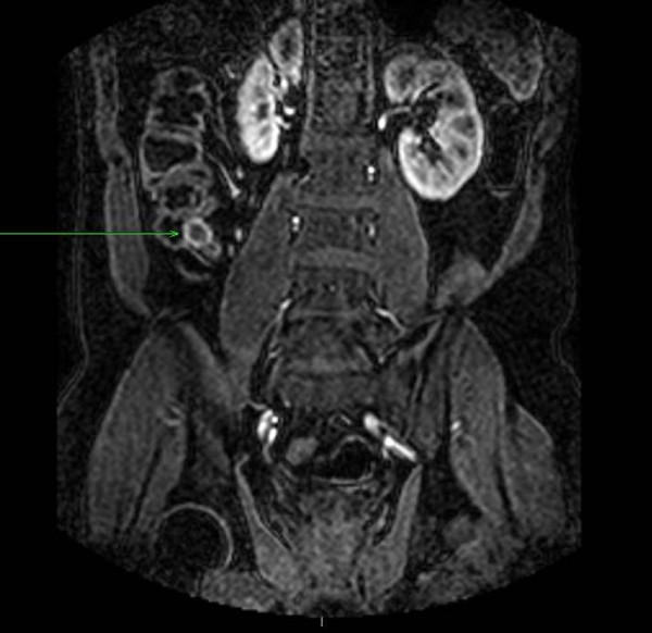
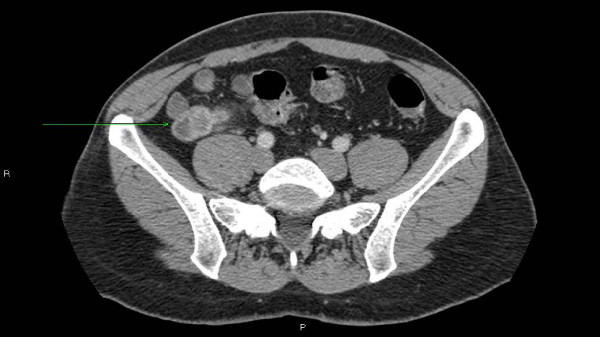
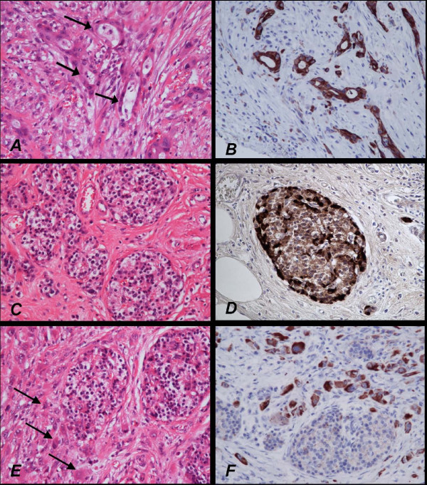
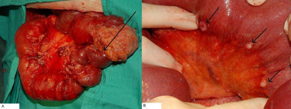
Case presentation: We describe a 50-year-old man who presented with rectal bleeding and anaemia, later found to be caused by a pancreatic adenocarcinoma arising from ectopic pancreatic tissue in a Meckel's diverticulum. The tumour was unfortunately highly aggressive, and the patient passed away within 5 months of symptom onset.
Conclusion: We believe this is the first case of pancreatic adenocarcinoma in a Meckel's diverticulum to be reported in the literature. The diagnosis of Meckel's should be considered in patients with acute gastrointestinal complaints; when found incidentally at laparotomy, it should be carefully examined for any gross abnormality and resection should be considered.
Figures
References
-
- Cates JM, Williams TL, Suriawinata AA. Intraductal papillary mucinous adenoma that arises from pancreatic heterotopia within a meckel diverticulum. Arch Pathol Lab Med . 2005;129:e67–e69. - PubMed
-
- Dolan RV, Remine WH, Dockerty MB. The fate of heterotopic pancreatic tissue – A study of 212 cases. Arch Surg. 1974;109:762–765. - PubMed
Publication types
MeSH terms
LinkOut - more resources
Full Text Sources
Medical
