

An MRI-based approach for the measurement of the dorsolateral prefrontal cortex in humans
- PMID: 19545981
- PMCID: PMC2778758
- DOI: 10.1016/j.pscychresns.2009.02.007
An MRI-based approach for the measurement of the dorsolateral prefrontal cortex in humans
Abstract
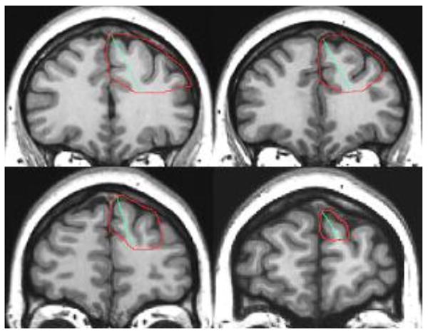
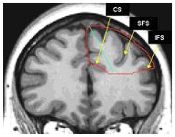
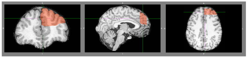
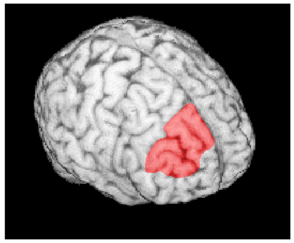
The dorsolateral prefrontal cortex (DLPFC) has been implicated in the pathophysiology of mental disorders. Previous region-of-interest MRI studies that attempted to delineate this region adopted various landmarks and measurement techniques, with inconsistent results. We developed a new region-of-interest measurement method to obtain morphometric data of this region from structural MRI scans, taking into account knowledge from cytoarchitectonic postmortem studies and the large inter-individual variability of this region. MRI scans of 10 subjects were obtained, and DLPFC tracing was performed in the coronal plane by two independent raters using the semi-automated software Brains2. The intra-class correlation coefficients between two independent raters were 0.94 for the left DLPFC and 0.93 for the right DLPFC. The mean +/- S.D. DLPFC volumes were 9.23 +/- 2.35 ml for the left hemisphere and 8.20 +/- 2.08 ml for the right hemisphere. Our proposed method has high inter-rater reliability and is easy to implement, permitting the standardized measurement of this region for clinical research applications
Figures
Similar articles
-
Inter-rater reliability of manual segmentation of the superior, inferior and middle frontal gyri.Psychiatry Res. 2006 Dec 1;148(2-3):151-63. doi: 10.1016/j.pscychresns.2006.05.006. Epub 2006 Nov 7. Psychiatry Res. 2006. PMID: 17088050
-
Definition of DLPFC and M1 according to anatomical landmarks for navigated brain stimulation: inter-rater reliability, accuracy, and influence of gender and age.Neuroimage. 2013 Sep;78:224-32. doi: 10.1016/j.neuroimage.2013.03.061. Epub 2013 Apr 6. Neuroimage. 2013. PMID: 23567888
-
Using 3D-MRI to localize the dorsolateral prefrontal cortex in TMS research.World J Biol Psychiatry. 2010 Mar;11(2 Pt 2):425-30. doi: 10.1080/15622970802669564. World J Biol Psychiatry. 2010. PMID: 19172531
-
Limits and reproducibility of resting-state functional MRI definition of DLPFC targets for neuromodulation.Brain Stimul. 2019 Jan-Feb;12(1):129-138. doi: 10.1016/j.brs.2018.10.004. Epub 2018 Oct 13. Brain Stimul. 2019. PMID: 30344110 Free PMC article.
-
Easy methods to make the neuronavigated targeting of DLPFC accurate and routinely accessible for rTMS.Neurophysiol Clin. 2017 Feb;47(1):35-46. doi: 10.1016/j.neucli.2017.01.007. Epub 2017 Feb 13. Neurophysiol Clin. 2017. PMID: 28202333
Cited by
-
Dorsolateral prefrontal cortex volume as a mediator between socioeconomic status and executive function.Neuropsychology. 2018 Nov;32(8):985-995. doi: 10.1037/neu0000484. Epub 2018 Sep 13. Neuropsychology. 2018. PMID: 30211609 Free PMC article.
-
Cigarette smoking and white matter microstructure in schizophrenia.Psychiatry Res. 2012 Feb 28;201(2):152-8. doi: 10.1016/j.pscychresns.2011.08.010. Epub 2012 Mar 3. Psychiatry Res. 2012. PMID: 22386966 Free PMC article.
-
Anhedonia correlates with functional connectivity of the nucleus accumbens subregions in patients with major depressive disorder.Neuroimage Clin. 2021;30:102599. doi: 10.1016/j.nicl.2021.102599. Epub 2021 Feb 23. Neuroimage Clin. 2021. PMID: 33662708 Free PMC article.
-
Mesoscale Brain Mapping: Bridging Scales and Modalities in Neuroimaging - A Symposium Review.Neuroinformatics. 2024 Oct;22(4):679-706. doi: 10.1007/s12021-024-09686-2. Epub 2024 Sep 23. Neuroinformatics. 2024. PMID: 39312131 Free PMC article. Review.
-
AGTR1 gene variation: association with depression and frontotemporal morphology.Psychiatry Res. 2012 May 31;202(2):104-9. doi: 10.1016/j.pscychresns.2012.03.007. Epub 2012 Jun 15. Psychiatry Res. 2012. PMID: 22703619 Free PMC article.
References
-
- Bearden CE, Hoffman KM, Cannon TD. The neuropsychology and neuroanatomy of bipolar affective disorder: a critical review. Bipolar Disorders. 2001;3:106–50. discussion 151-3. - PubMed
-
- Bench CJ, Frackowiak RS, Dolan RJ. Changes in regional cerebral blood flow on recovery from depression. Psychological Medicine. 1995;25:247–61. - PubMed
-
- Bunney WE, Bunney BG. Evidence for a compromised dorsolateral prefrontal cortical parallel circuit in schizophrenia. Brain Research Brain Research Reviews. 2000;31:138–46. - PubMed
-
- Cotter D, Mackay D, Chana G, Beasley C, Landau S, Everall IP. Reduced neuronal size and glial cell density in area 9 of the dorsolateral prefrontal cortex in subjects with major depressive disorder. Cereb Cortex. 2002;12(4):386–94. - PubMed
-
- Crespo-Facorro B, Kim JJ, Andreasen NC, O'Leary DS, Wiser AK, Bailey JM, Harris G, Magnotta VA. Human frontal cortex: an MRI-based parcellation method. Neuroimage. 1999;10:500–19. - PubMed
Publication types
MeSH terms
Grants and funding
LinkOut - more resources
Full Text Sources
Medical
