Randomized Controlled Trial
. 2009 Aug 1;587(Pt 15):3869-83.
doi: 10.1113/jphysiol.2009.169425. Epub 2009 Jun 22.

Electrophysiological properties of thalamic, subthalamic and nigral neurons during the anti-parkinsonian placebo response

Affiliations
Randomized Controlled Trial

Electrophysiological properties of thalamic, subthalamic and nigral neurons during the anti-parkinsonian placebo response

Fabrizio Benedetti et al. J Physiol. .

Abstract

Placebo administration to Parkinson patients is known to induce dopamine release in the striatum and to affect the activity of subthalamic nucleus (STN) neurons. By using intraoperative single-neuron recording techniques in awake patients, here we extend our previous study on STN recording, and characterize part of the neuronal circuit which is affected by placebos. In those patients who showed a clinical placebo response, there was a decrease in firing rate in STN neurons that was associated with a decrease in the substantia nigra pars reticulata (SNr) and an increase in the ventral anterior (VA) and anterior ventral lateral (VLa) thalamus. These data show that placebo decreases STN and SNr activity whereas it increases VA/VLa activity. By contrast, placebo non-responders showed either a lack of changes in this circuit or partial changes in the STN only. Thus, changes in activity in the whole basal ganglia-VA/VLa circuit appear to be important in order to observe a clinical placebo improvement, although the involvement of other circuits, such as the direct pathway bypassing the STN, cannot be ruled out. The circuit we describe in the present study is likely to be a part of a more complex circuitry, including the striatum and the internal globus pallidus (GPi), that is modified by placebo administration. These findings indicate that a placebo treatment, which is basically characterized by verbal suggestions of benefit, can reverse the malfunction of a complex neuronal circuit, although these placebo-associated neuronal changes are short-lasting and occur only in some patients but not in others.


Figures

Figure 1
Figure 1. The neuronal circuit analysed in this study
A, the circles represent the recorded neurons. The subthalamic nucleus (STN) neurons, which receive inputs from the cortex, the striatum, the external globus pallidus (GPe) as well as from other regions, send their output excitatory information to different regions, such as the substantia nigra pars reticulata (SNr) and the internal globus pallidus (GPi). SNr has an inhibitory connection with the thalamus, and the thalamus sends projections to the motor cortex. The striatum also sends projections to GPi, which in turn projects to the thalamus, and to SNr. B, magnetic resonance imaging of the electrode track with the electrode tip in the thalamic–subthalamic region. The square represents the region which is magnified in C. C, magnification of the square in B. It can be seen that the electrode track passes through VA, VLa, STN and SNr. We could record from all these regions during the placebo response, thus analysing the circuit shown in A.
Figure 2
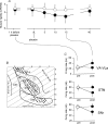
Figure 2. Data from all the patients who received the placebo treatment and from those who received no treatment (mean ±s.d.)
A, the clinical placebo response (filled circles) is compared with the no-treatment group (open circles). Pre-placebo recordings were performed 1 h before placebo treatment, whereas post-placebo recordings were carried out starting from 15 min (maximum of the response) after placebo administration. B, location of the recorded neurons on the Schaltenbrand and Wahren atlas (Schaltenbrand & Wahren, 1977). It is important to note that many recording sites overlap, so that their number turns out to be smaller than the actual number of recorded units. C, neuronal firing rate in VA/VLa, STN and SNr, before (open circles) and after (filled circles) placebo (continuous lines). The dashed lines show the firing rate in the no-treatment group on the first side (open circles) and second side (filled circles) of recording. Note that during the maximum placebo response, VA/VLa neuronal activity increased whereas STN and SNr activity decreased.
Figure 3
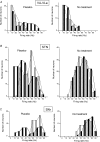
Figure 3. Distribution of the frequencies in the placebo group (left) and the no-treatment group (right) in VA/VLa (A), STN (B) and SNr (C)
On the left, the shaded bars and dashed line show the pre-placebo condition whereas the black bars and the continuous line show the post-placebo condition. On the right, the shaded bars and the dashed line show the first recording side whereas the filled bars and the continuous line show the second recording side. Note the increased frequencies in VA/VLa and the decreased frequencies in STN and SNr after placebo. No changes are present in the no-treatment group.
Figure 5
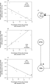
Figure 5. Correlation between the percentage of neuronal activity change of STN and that of VA/VLa (A), STN and SNr (B), SNr and VA/VLa (C)
The pattern of correlation, positive in B and negative in A and C, supports the excitatory connection between STN and SNr, and the inhibitory connection between SNr and VA/VLa (D).
Figure 4
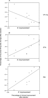
Figure 4. Correlation between percentage of clinical improvement and percentage of neuronal activity change of VA/VLa (A), STN (B) and SNr (C)
In all cases there was a high correlation, according to the following rule: the larger the clinical improvement, the lower the firing rate in STN and SNr and the higher the firing rate in VA/VLa.
Figure 6
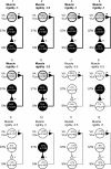
Figure 6. Deactivation (black) and activation (grey) pattern of the STN–SNr–VA/VLa circuit in placebo responders (subjects 1–6) and non-responders (subjects 7–12)
The percentage decrease or increase in neuronal activity after placebo administration is shown along with statistical significance. The UPDRS decrease in muscle rigidity after placebo (clinical placebo response) is also shown. Note that STN and SNr are deactivated and VA/VLa is activated only in those subjects with a reduction in muscle rigidity equal to or larger than 1 UPDRS (responders). By contrast, no neuronal changes were present (white neurons) in those subjects with muscle rigidity reduction smaller than 1 UPDRS (non-responders). Also note that clinical non-responders 8 and 10 showed only partial changes, with a significant deactivation of STN but no changes in SNr and VA/VLa.

Similar articles

Cited by

References

    1. Albin RL, Young AB, Penney JB. The functional anatomy of basal ganglia disorders. Trends Neurosci. 1989;12:366–375. - PubMed
    1. Amanzio M, Benedetti F. Neuropharmacological dissection of placebo analgesia: expectation activated opioid systems versus conditioning-activated specific subsystems. J Neurosci. 1999;19:484–494. - PMC - PubMed
    1. Benarroch EE. Subthalamic nucleus and its connections: Anatomic substrate for the network effects of deep brain stimulation. Neurology. 2008;70:1991–1995. - PubMed
    1. Benazzouz A, Gao DM, Ni ZG, Piallat B, Bouali-Benazzouz R, Benabid AL. Effect of high-frequency stimulation of the subthalamic nucleus on the neuronal activities of the substantia nigra pars reticulata and ventrolateral nucleus of the thalamus in the rat. Neuroscience. 2000;99:289–295. - PubMed
    1. Benazzouz A, Hallett M. Mechanism of action of deep brain stimulation. Neurology. 2000;55(Suppl. 6):S13–S17. - PubMed

Publication types

MeSH terms

Substances