. 2009 Aug 21;284(34):23046-58.
doi: 10.1074/jbc.M109.032631. Epub 2009 Jun 22.

Hypoxia-inducible factor prolyl-4-hydroxylase PHD2 protein abundance depends on integral membrane anchoring of FKBP38

Affiliations

Hypoxia-inducible factor prolyl-4-hydroxylase PHD2 protein abundance depends on integral membrane anchoring of FKBP38

Sandra Barth et al. J Biol Chem. .

Abstract

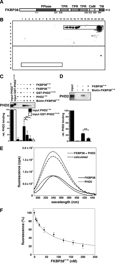
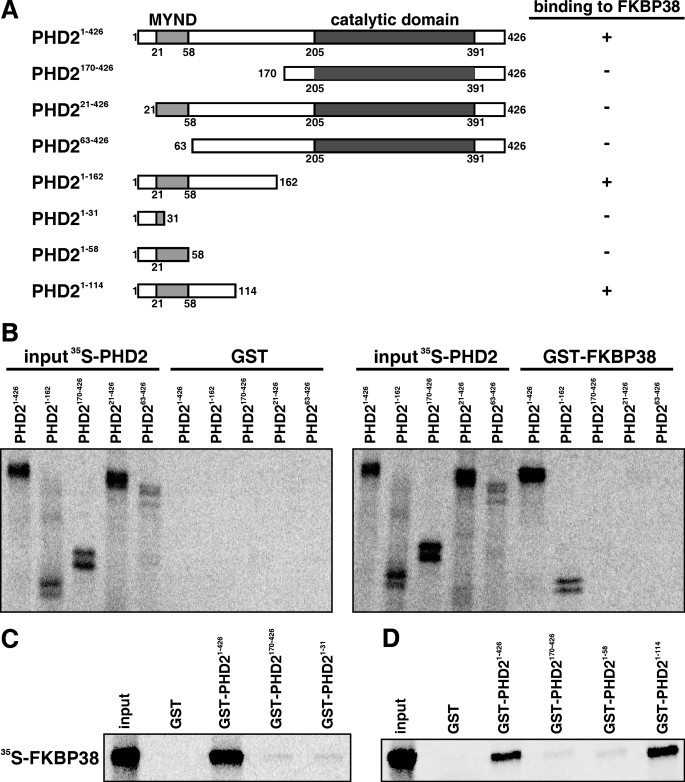
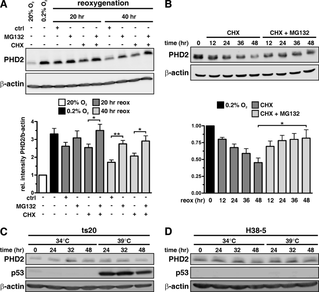
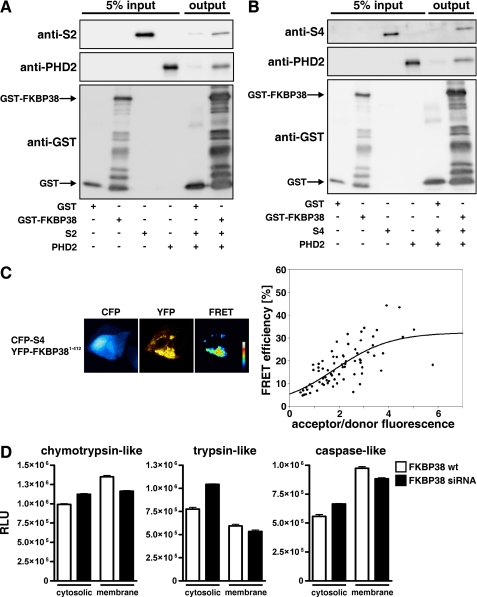
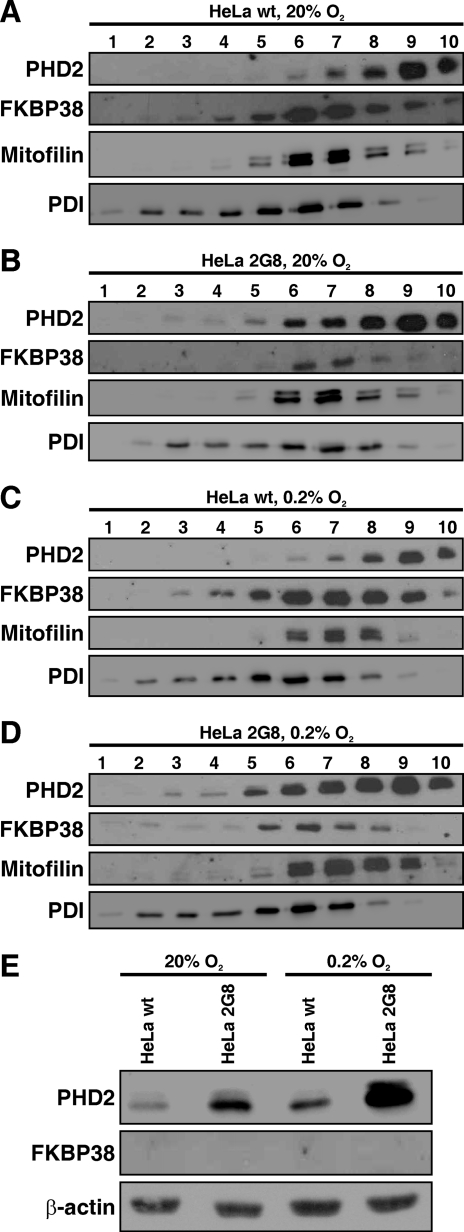
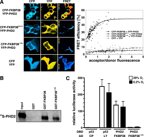
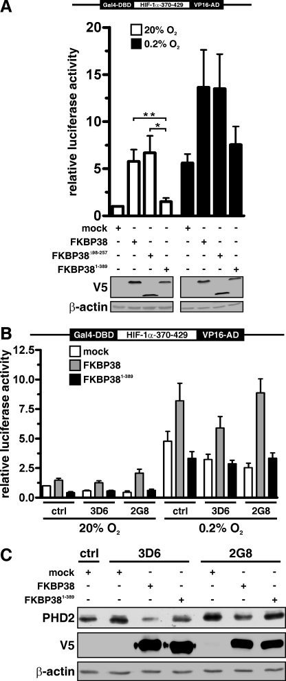
Prolyl-4-hydroxylase domain (PHD) proteins are 2-oxoglutarate and dioxygen-dependent enzymes that mediate the rapid destruction of hypoxia-inducible factor alpha subunits. Whereas PHD1 and PHD3 proteolysis has been shown to be regulated by Siah2 ubiquitin E3 ligase-mediated polyubiquitylation and proteasomal destruction, protein regulation of the main oxygen sensor responsible for hypoxia-inducible factor alpha regulation, PHD2, remained unknown. We recently reported that the FK506-binding protein (FKBP) 38 specifically interacts with PHD2 and determines PHD2 protein stability in a peptidyl-prolyl cis-trans isomerase-independent manner. Using peptide array binding assays, fluorescence spectroscopy, and fluorescence resonance energy transfer analysis, we defined a minimal linear glutamate-rich PHD2 binding domain in the N-terminal part of FKBP38 and showed that this domain forms a high affinity complex with PHD2. Vice versa, PHD2 interacted with a non-linear N-terminal motif containing the MYND (myeloid, Nervy, and DEAF-1)-type Zn(2+) finger domain with FKBP38. Biochemical fractionation and immunofluorescence analysis demonstrated that PHD2 subcellular localization overlapped with FKBP38 in the endoplasmic reticulum and mitochondria. An additional fraction of PHD2 was found in the cytoplasm. In cellulo PHD2/FKBP38 association, as well as regulation of PHD2 protein abundance by FKBP38, is dependent on membrane- anchored FKBP38 localization mediated by the C-terminal transmembrane domain. Mechanistically our data indicate that PHD2 protein stability is regulated by a ubiquitin-independent proteasomal pathway involving FKBP38 as adaptor protein that mediates proteasomal interaction. We hypothesize that FKBP38-bound PHD2 is constantly degraded whereas cytosolic PHD2 is stable and able to function as an active prolyl-4-hydroxylase.


Figures

FIGURE 1.
FIGURE 1.
Analysis of the PHD2 interaction domain in FKBP38. A, domain architecture of FKBP381–412. PPIase, peptidyl-prolyl cis-trans isomerase; TPR, tetratricopeptide repeats; CaM, calmodulin-binding site; TM, TM domain. B, an array of 13-mer peptides spanning the FKBP38 sequence was synthesized with forward shifts by 1 amino acid. PHD2 interaction with the peptide array of the FKBP38 forward sequence (upper panel) and reverse sequence (lower panel) was analyzed by immunoblotting. The PHD2 interaction pattern is displayed in the framed region. The respective binding motif comprised the peptides b13–24, which correspond to FKBP3837–56. The reverse sequence comprising the same residues in the reverse order in the peptides f1–12 was not found to interact with PHD2 (lower panel). C, immunoblot analysis of the interaction between two different PHD2 variants and a biotin-labeled FKBP3837–56 peptide-bound streptavidin matrix using anti-PHD2 antibodies. The streptavidin matrix alone served as control. D, endogenous proteins from rat liver lysates were incubated with an FKBP3837–56 affinity matrix, and PHD2 binding was detected by immunoblotting. The streptavidin matrix alone served as binding control. Representative values in the lower column diagrams are average relative band intensities with S.E. of several independent experiments. p values were obtained by unpaired t tests (*, p < 0.05; **, p < 0.01). E, fluorescence measurements at an excitation wavelength of 278 nm with 1 μm FKBP38 (—), 1 μm PHD2 (····), and a 1:1 mixture of both proteins (- - - -). The calculated spectrum (_ _ _) represents the sum of the individual protein spectra as it should appear when the components do not interact. F, titration curve resulting from fluorescence measurements at 340 nm (excitation at 278 nm) of a sample containing 1 μm FKBP38/PHD2 and various concentrations of a peptide corresponding to FKBP3837–56. The straight line represents the fit according to the equation under “Experimental Procedures.” rel., relative; cps, counts/s.
FIGURE 2.
FIGURE 2.
Mapping the interaction domain of PHD2. A, schematic representation of the PHD2 domain architecture and the PHD2 constructs used. B, IVTT 35S-labeled PHD2 variants were incubated with GST-FKBP38 or GST alone. Protein complexes were pulled down with glutathione-Sepharose beads, separated by SDS-PAGE, and visualized by phosphorimaging. C and D, IVTT 35S-labeled FKBP38 was incubated with recombinant GST-PHD21–426, GST-PHD2170–426, GST-PHD21–31, GST-PHD21–58, GST-PHD21–114, or GST alone. Protein complexes were pulled down and visualized as described above.
FIGURE 3.
FIGURE 3.
Proteolytic regulation of PHD2. Cellular extracts were prepared, separated by SDS-PAGE, and analyzed by immunoblotting. A, MCF-7 cells were preincubated for 24 h at 0.2% O2 and then reoxygenated for 20 or 40 h in the presence of solvent control, MG132 (5 μm), and/or CHX (50 μm). B, MCF-7 cells were cultivated for 24 h under 0.2% O2 before CHX (50 μm) and solvent control or CHX and MG132 (5 μm) were added for the indicated time periods. PHD2 and β-actin protein levels were analyzed by immunoblotting. C, mouse ts20 cells were cultivated at either 34 or 39 °C for 24, 32, or 48 h, and cellular extracts were analyzed by immunoblotting. D, mouse ts20 cells reconstituted with a wild-type E1 gene (H38-5) were incubated under the same conditions as described in C, and cellular proteins were analyzed by immunoblotting. reox, reoxygenation; ctrl, control; rel., relative. *, p < 0.05; **, p < 0.01.
FIGURE 4.
FIGURE 4.
FKBP38 regulates proteasomal activity. A and B, recombinant GST or GST-FKBP38 proteins were incubated with recombinant S2 or S4 and PHD2 proteins, and protein complexes were pulled down with glutathione-Sepharose beads, separated by SDS-PAGE, and visualized by immunoblotting. C, HEK293 cells were transiently transfected with CFP-S4 and YFP-FKBP38 plasmids, and FRET analysis was performed. Subcellular distribution of FRET efficiency signals ranging from 0 to 60% was visualized in false color mode as indicated by the color bar (black, 0%; white, 60%). FRET efficiencies of single cells were averaged and plotted to the acceptor/donor fluorescence ratio. D, cytosolic or membrane fractions of parental or FKBP38-down-regulated 2G8 HeLa cells were incubated with Suc-LLVY-, Z-LRR-, or Z-nLPnLD-aminoluciferin. Results are mean values ±S.E. of n = 3 independent experiments performed in triplicates. wt, wild type; RLU, relative luciferase units.
FIGURE 5.
FIGURE 5.
Analysis of FKBP38 and PHD2 protein levels by biochemical fractionation. Parental HeLa (wild type (wt)) or FKBP38-silenced (2G8) HeLa cells were cultivated at 20% O2 (A and B) or at 0.2% O2 (C and D). Cellular membranes were separated from cytosolic fractions by differential centrifugation and then separated in a 10–30% iodixanol gradient. 1-ml fractions were collected and analyzed by immunoblotting. Mitofilin served as a mitochondria marker, and protein-disulfide isomerase (PDI) served as an ER marker. E, cytosolic fractions of parental HeLa and HeLa 2G8 cells were analyzed by immunoblotting.
FIGURE 6.
FIGURE 6.
FKBP38 transmembrane domain is required for PHD2 interaction. A, HEK293 cells were transiently transfected with the indicated CFP or YFP plasmids, and FRET analysis was performed. B, recombinant GST, GST-FKBP38, or GST-FKBP381–389 proteins were incubated with IVTT 35S-labeled PHD2, and protein complexes were pulled down with glutathione-Sepharose beads, separated by SDS-PAGE, and visualized by phosphorimaging. C, HeLa cells were transiently transfected with Gal4 DBD and Gal4 activation domain (VP16-AD) fusion protein vectors, Gal4 response element-driven firefly luciferase reporter, and a Renilla luciferase control vector. Following transfection, the cells were incubated under normoxic (20% O2) or hypoxic (0.2% O2) conditions, and luciferase reporter gene activities were determined 16 h later. Firefly to Renilla luciferase activity ratios were normalized to the normoxic negative control DBD-p53/AD-CP (CP) (VP3 polyoma virus coat protein) co-transfection that was arbitrarily defined as 1. DBD-p53/AD-LT (LT) (large T antigen) served as a positive control. Mean values ±S.E. are shown of n = 3 independent experiments performed in triplicates.
FIGURE 7.
FIGURE 7.
FKBP38 transmembrane domain is necessary for functional regulation of PHD2. A, MCF-7 cells were transiently transfected with Gal4-DBD-HIF-1α370–429-VP16-AD expression vector, Gal4 response element-driven firefly luciferase reporter, and a Renilla luciferase control vector and either co-transfected with V5-FKBP38, V5-FKBP38Δ98–357, V5-FKBP381–389, or a mock plasmid (pcDNA3.1-LacZ). 24 h post-transfection, cells were either cultured under normoxic or hypoxic conditions for an additional 16 h before relative luciferase activities were determined. Results are presented as mean values of relative luciferase activities ±S.E. of n = 4 independent experiments performed in triplicates. p values were obtained by paired t tests (**, p < 0.01; *, p < 0.05). Expression of the transfected vectors was verified by immunoblotting against the V5 tag. B, HeLa FKBP38 RNAi control cells (ctrl) and FKBP38 RNAi-depleted cells (3D6 and 2G8) were transiently transfected with the indicated plasmids as described in A. Results are mean values ±S.E. of n = 7 independent experiments performed in triplicates. C, parental HeLa as well as RNAi control (ctrl) and FKBP38 down-regulated (3D6 and 2G8) cells were transiently transfected with the indicated plasmids, and PHD2, V5, and β-actin were detected by immunoblotting.

References

    1. Semenza G. L. (2007) Sci. STKE 2007,cm8. - PubMed
    1. Wenger R. H., Stiehl D. P., Camenisch G. (2005) Sci. STKE 2005,re12. - PubMed
    1. Bruick R. K., McKnight S. L. (2001) Science 294,1337–1340 - PubMed
    1. Epstein A. C., Gleadle J. M., McNeill L. A., Hewitson K. S., O'Rourke J., Mole D. R., Mukherji M., Metzen E., Wilson M. I., Dhanda A., Tian Y. M., Masson N., Hamilton D. L., Jaakkola P., Barstead R., Hodgkin J., Maxwell P. H., Pugh C. W., Schofield C. J., Ratcliffe P. J. (2001) Cell 107,43–54 - PubMed
    1. Ivan M., Haberberger T., Gervasi D. C., Michelson K. S., Günzler V., Kondo K., Yang H., Sorokina I., Conaway R. C., Conaway J. W., Kaelin W. G., Jr. (2002) Proc. Natl. Acad. Sci. U.S.A. 99,13459–13464 - PMC - PubMed

Publication types

MeSH terms

Substances

LinkOut - more resources