. 2009 Sep;29(17):4595-603.
doi: 10.1128/MCB.00275-09. Epub 2009 Jun 22.

A randomly integrated transgenic H19 imprinting control region acquires methylation imprinting independently of its establishment in germ cells

Affiliations

A randomly integrated transgenic H19 imprinting control region acquires methylation imprinting independently of its establishment in germ cells

Hitomi Matsuzaki et al. Mol Cell Biol. 2009 Sep.

Abstract

The imprinted expression of the mouse Igf2/H19 locus is governed by the differential methylation of the imprinting control region (ICR), which is established initially in germ cells and subsequently maintained in somatic cells, depending on its parental origin. By grafting a 2.9-kbp H19 ICR fragment into a human beta-globin yeast artificial chromosome in transgenic mice, we previously showed that the ICR could recapitulate imprinted methylation and expression at a heterologous locus, suggesting that the H19 ICR in the beta-globin locus contained sufficient information to maintain the methylation mark (K. Tanimoto, M. Shimotsuma, H. Matsuzaki, A. Omori, J. Bungert, J. D. Engel, and A. Fukamizu, Proc. Natl. Acad. Sci. USA 102:10250-10255, 2005). Curiously, however, the transgenic H19 ICR was not methylated in sperm, which was distinct from that seen in the endogenous locus. Here, we reevaluated the ability of the H19 ICR to mark the parental origin using more rigid criteria. In the testis, the methylation levels of the solitary 2.9-kbp transgenic ICR fragment varied significantly between six transgenic mouse lines. However, in somatic cells, the paternally inherited ICR fragment exhibited consistently higher methylation levels at five out of six randomly integrated sites in the mouse genome. These results clearly demonstrated that the H19 ICR could acquire parent-of-origin-dependent methylation after fertilization independently of the chromosomal integration site or the prerequisite methylation acquisition in male germ cells.


Figures

FIG. 1.
FIG. 1.
Generation and characterization of H19 ICR TgM. (A) Genomic structure of the mouse Igf2/H19 locus. The H19 ICR (DMD) is located within a 2.9-kbp SacI (Sa; at −4.7 kbp relative to the transcription initiation site of the H19 gene)-BamHI (B; at −1.8 kbp) fragment. The solid rectangles in the enlarged map indicate the position of the CTCF binding sites. (B) Schematic representation of the transgenes. In ICR/β-globin (top; previously referred to as ICR), the 2.9-kbp ICR fragment (inverted orientation) was floxed (filled triangles) and introduced between HS1 and the ɛ-globin gene in the human β-globin YAC (32). In the HS1/ICR transgene (bottom), the 2.9-kbp ICR fragment was flanked by loxP (filled triangles) and ∼0.5-kbp human β-globin locus sequences (gray boxes) on both sides, which are the same as those surrounding the ICR fragment in the ICR/β-globin YAC. (C) Copy number analysis of the HS1/ICR transgenes. Genomic DNA was digested with BamHI, and blots were hybridized with the BS probe (see Fig. 2A). The copy numbers of the HS1/ICR transgene in six established TgM lines were determined by comparing the transgenic (Tg) ICR signals to that of ICR/β-globin YAC TgM (line 1048; single copy) after normalization to the endogenous (endo) ICR signals.
FIG. 2.
FIG. 2.
Methylation of the ICR in erythroid cells. (A) Restriction map of the endogenous H19 (top) and the HS1/ICR transgene (bottom) loci. Methylation-sensitive HhaI sites in the BamHI fragments (horizontal lines beneath each map) within the locus are displayed as vertical lines. Fragments from the endogenous and transgenic H19 ICR could be distinguished by their sizes because of the transgene-specific BamHI site. A probe (BS) used for Southern blot analyses is shown as a filled rectangle. B, BamHI; E, EcoRI; Sa, SacI. (B) Genomic DNA from nucleated erythroid cells was prepared from TgM that inherited the transgenes either paternally (P) or maternally (M) or from nontransgenic (non-Tg) mice. DNA was digested with BamHI (−) and then HhaI (Hh) and analyzed by Southern blotting with the BS probe. endo, endogenous H19 ICR; Tg, the transgenic H19 ICR. Asterisks indicate the positions of parental or methylated, undigested fragments. (C) Methylation analysis by bisulfite sequencing. (Top) Two distinct regions (I and II) of the transgenic ICR were amplified by nested PCR. The positions of the primers are shown by arrows. Each one of the primers for first-round PCRs was set at the β-globin sequences in the transgene. (Bottom) Genomic DNA from nucleated erythroid cells (line 362) was prepared from male (upper) or female (lower) mice, which inherited the transgene either paternally (P) or maternally (M), and was analyzed by bisulfite sequencing. Each horizontal row represents a single DNA template molecule. Methylated (filled circles) and unmethylated (open circles) CpG motifs are shown. Gray bars indicate the location of the CTCF binding sites.
FIG. 3.
FIG. 3.
Methylation of the ICR in testis. (A) Genomic DNA was prepared from the testis of TgM that inherited the transgenes either paternally (P) or maternally (M) or from nontransgenic (non-Tg) mice. DNA was digested with BamHI (−) and then HhaI (Hh) and analyzed by Southern blotting with the BS probe. endo, endogenous H19 ICR; Tg, transgenic H19 ICR. Asterisks indicate the positions of the parental or methylated, undigested fragments. (B) Genomic DNA was extracted from the testis of TgM (line 362) that had inherited the transgene either paternally (P) or maternally (M). The methylation status of the transgenic H19 ICR was analyzed by bisulfite sequencing as described in the legend to Fig. 2C. (C) Copy number analysis of the Cre-deleted TgM sublines. Tail DNA was digested with BamHI, and the blot was hybridized with the BS probe. DNA from TgM of the parental five-copy line 378 and the low-copy-number sublines (four copies and single copy) obtained by in vivo Cre-loxP recombination was analyzed. The copy numbers of each line were determined as described in Materials and Methods. (D) Genomic DNA was extracted from the testis of low-copy-number sublines of line 378, inheriting the transgene either paternally (P) or maternally (M). DNA was digested with BamHI and then HhaI, size fractionated on the agarose gel, transferred to nylon membranes, and hybridized with the BS probe. In all experiments, testis was recovered from male animals inheriting transgenes either maternally or paternally for monitoring the contamination of somatic cells in the samples. Since differences in the two groups were minimal, we presumed that the methylation pattern of the transgenic ICR in the whole testis was represented mostly by germ cells in the testis.
FIG. 4.
FIG. 4.
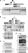
Reprogramming of DNA methylation in the Cre-deleted transgenic ICR. (A and B, top) The pedigrees of the single-copy (A) or the four-copy (B) sublines from line 378. Hemizygous male and female TgM are represented by filled squares and open circles, respectively. Only the animals analyzed in this experiment are shown. (A and B, bottom) Tail DNA from TgM shown in the pedigrees was digested with BamHI and then HhaI. Blots were hybridized with the BS probe. The numbers above each panel correspond to those in the pedigrees. (C and D) DNA methylation status of the transgenic ICR (region II) in oocytes. Oocyte DNA from the single-copy (C) and four-copy (D) sublines of TgM, which inherited the transgenes either paternally (P) or maternally (M), was analyzed by bisulfite sequencing. The results from each independent reaction are separately presented by the gap between clusters. The numbers to the left of each row indicate the number of times the pattern was observed in sequenced PCR products. Some oocytes were collected from the females shown in the pedigrees in panels A and B, as indicated to the right of each row. In the experiment using oocytes from the four-copy TgM that inherited the transgene paternally (D, top), second-round PCR, subcloning, and sequencing were performed twice to eliminate cloning bias.
FIG. 5.
FIG. 5.
Methylation of injected ICR fragments in embryos. The methylation-free HS1/ICR DNA fragment was injected (inj.) into the male or female pronuclei of fertilized eggs. Eggs were transferred into the oviducts of foster mothers. At embryonic day 10.5, embryos were recovered and genotyped for the transgenic ICR. Fourteen embryos from three independent injections carried the injected ICR. Genomic DNA of the embryos was treated with sodium bisulfite, and the transgenic ICR regions II (A) and I (B) were amplified by nested PCR (Fig. 2C). The aliquots of PCR products were digested (+) with TaqI to assess the methylation status of the original DNA. The effectiveness of bisulfite mutagenesis was validated by the digestion of the PCR products with MseI (data not shown). The appearance of the smaller fragments represents the hypermethylation of the ICR at the enzyme recognition sites. TaqI sites (vertical lines) in the PCR products (horizontal lines) and expected restriction fragment sizes are shown below each panel.

References

    1. Ainscough, J. F., T. Koide, M. Tada, S. Barton, and M. A. Surani. 1997. Imprinting of Igf2 and H19 from a 130 kb YAC transgene. Development 1243621-3632. - PubMed
    1. Bartolomei, M. S., S. Zemel, and S. M. Tilghman. 1991. Parental imprinting of the mouse H19 gene. Nature 351153-155. - PubMed
    1. Bell, A. C., and G. Felsenfeld. 2000. Methylation of a CTCF-dependent boundary controls imprinted expression of the Igf2 gene. Nature 405482-485. - PubMed
    1. Birger, Y., R. Shemer, J. Perk, and A. Razin. 1999. The imprinting box of the mouse Igf2r gene. Nature 39784-88. - PubMed
    1. Bowman, A. B., J. M. Levorse, R. S. Ingram, and S. M. Tilghman. 2003. Functional characterization of a testis-specific DNA binding activity at the H19/Igf2 imprinting control region. Mol. Cell. Biol. 238345-8351. - PMC - PubMed

Publication types

LinkOut - more resources