. 2009 Nov;57(11):1013-23.
doi: 10.1369/jhc.2009.953539. Epub 2009 Jun 22.

Toll-like receptors 3, 4, and 7 are expressed in the enteric nervous system and dorsal root ganglia

Affiliations

Toll-like receptors 3, 4, and 7 are expressed in the enteric nervous system and dorsal root ganglia

Isabella Barajon et al. J Histochem Cytochem. 2009 Nov.

Abstract

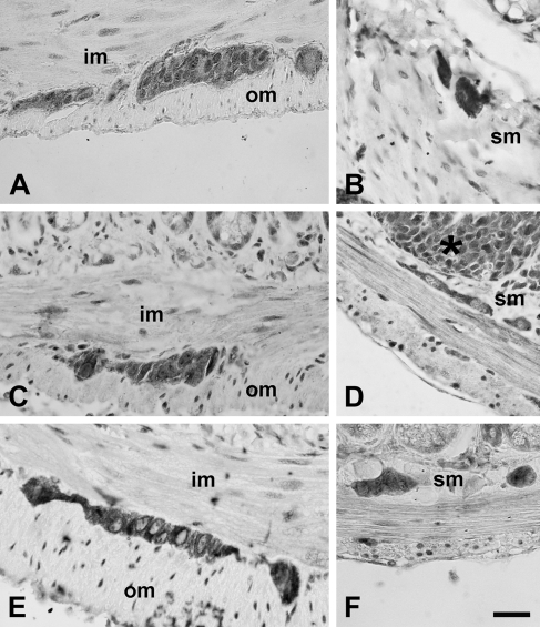
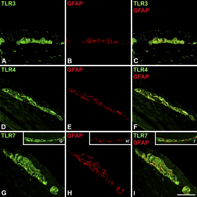
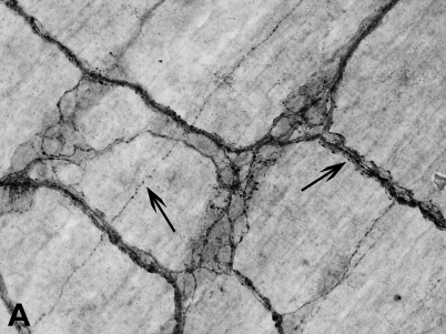
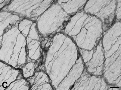
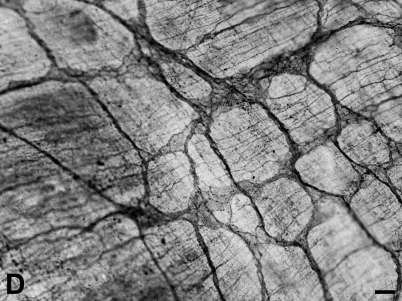
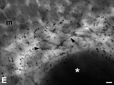
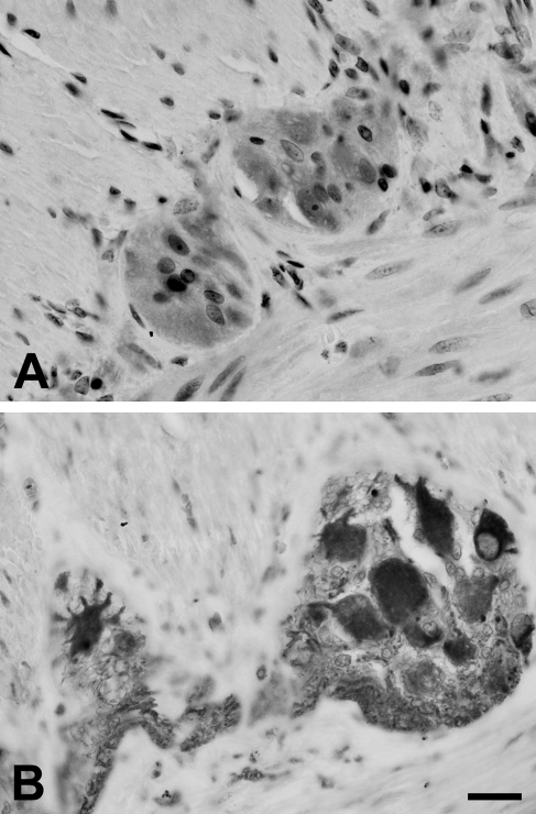
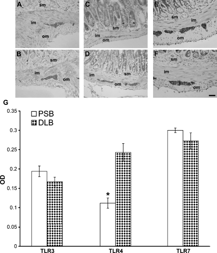
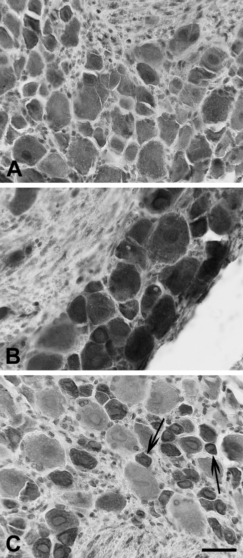
The aim of the present study was to evaluate the expression of innate immunity receptors belonging to the Toll-like family in the neural plexuses of the different tracts of murine intestine, of the human ileum, and in lower dorsal root ganglia (DRGs) from where extrinsic afferents to these plexuses originate. Results obtained by immunohistochemistry and immunofluorescence on paraffin-embedded tissue and whole-mount preparations show that Toll-like receptors (TLRs) -3 and -7, recognizing viral RNA, and TLR4, recognizing lipopolysaccharide (membrane component of Gram-negative bacteria), are expressed in the myenteric and submucous plexuses of murine intestine and human ileum, and in DRGs primary sensory neurons. They also show that TLR4 immunostaining is stronger in murine distal large bowel. In murine tissue, expression of TLRs was present in both neurons and glial cells. These observations indicate that the enteric neural network might be directly activated by bacterial and viral components and is therefore more in the forefront than previously envisaged in defense responses of the intestinal wall and in the cross-talk with intestinal microbiota. They also highlight the presence of a peripheral neural network that by way of hardwired neurotransmission could potentially convey to the central nervous system specific information on our microbial counterpart and invading or potentially invading pathogens.


Figures

Figure 1
Figure 1
Immunohistochemical labeling for Toll-like receptor 3 (TLR3) (A,B), TLR4 (C,D), and TLR7 (E,F) in the myenteric plexus (left column) and in the submucous plexus (right column) of murine intestine. im, inner muscle layer; om, outer muscle layer; sm, submucosa. Asterisk, Peyer's patch. Bar = 30 μm.
Figure 2
Figure 2
Confocal scanning microscopy analysis of TLR3 (A), TLR4 (D), TLR7 (G), and glial fibrillary acidic protein (GFAP) (B,E,H) in murine myenteric plexus. For each antigen, a single optical confocal section is shown. Merged images (C,F,I) evidence colocalization of TLRs and GFAP (yellow). In the inserts (G′,H′,I′), a plexus where glial cells are negative for TLR7 is shown. Bar = 30 μm.
Figure 3
Figure 3
Whole-mount immunohistochemistry of murine small bowel. (A–C) Whole-mount preparations of murine myenteric plexus and outer muscle layer showing ganglia and strands of nerve fibers positive for TLR3 (A), TLR4 (B), and TLR7 (C). (A) Arrows indicate nerve fibers with varicosities. In the right column, two examples of whole-mount preparations of the submucous plexus and inner smooth muscle layer are shown. (D) Immunostaining for TLR3. (E) TLR7-immunopositive submucous plexus (arrowheads) surrounding a Peyer's patch (asterisk). m, mucosa where flattened villi are visiible. Bars: C,D = 30 μm; E = 120 μm.
Figure 3
Figure 3
Whole-mount immunohistochemistry of murine small bowel. (A–C) Whole-mount preparations of murine myenteric plexus and outer muscle layer showing ganglia and strands of nerve fibers positive for TLR3 (A), TLR4 (B), and TLR7 (C). (A) Arrows indicate nerve fibers with varicosities. In the right column, two examples of whole-mount preparations of the submucous plexus and inner smooth muscle layer are shown. (D) Immunostaining for TLR3. (E) TLR7-immunopositive submucous plexus (arrowheads) surrounding a Peyer's patch (asterisk). m, mucosa where flattened villi are visiible. Bars: C,D = 30 μm; E = 120 μm.
Figure 4
Figure 4
Immunolabeling for TLR3 (A) and TLR7 (B) in the myenteric plexus of the human intestine. Bar = 30 μm.
Figure 5
Figure 5
Immunolabeling for TLR3 (A,B), TLR4 (C,D), and TLR7 (E,F) in murine proximal small bowel (upper row) and distal large bowel (lower row). TLR4 immunostaining appears to be stronger in the distal large bowel (D). im, inner muscle layer; om, outer muscle layer; sm, submucosa. Bar = 30 μm. (G) Quantitative analysis of TLR3, TLR4, and TLR7 immunostaining in proximal small bowel (PSB) and distal large bowel (DLB), determined by optical density (OD). Values are presented as mean ± SEM. Statistically significant difference was reached only for TLR4. *p<0.01.
Figure 6
Figure 6
Immunolabeling for TLR3 (A), TLR4 (B), and TLR7 (C) in murine dorsal root ganglia (DRGs). In C, arrows indicate small-sized neurons strongly reactive for TLR7. Bar = 30 μm.
Figure 7
Figure 7
Western blot analysis of protein extract from mouse lower thoracic and lumbosacral DRGs, probed with the antibodies anti-TLR4 (A), anti-TLR3 (B), and anti-TLR7 (C). Spleen protein extract was used as positive control.

Similar articles

Cited by

References

    1. Akira S, Uematsu S, Takeuchi O (2006) Pathogen recognition and innate immunity. Cell 124:783–801 - PubMed
    1. Berthoud HR, Blackshaw LA, Brookes SJ, Grundy D (2004) Neuroanatomy of extrinsic afferents supplying the gastrointestinal tract. Neurogastroenterol Motil 16(suppl 1):28–33 - PubMed
    1. Bomsel M, Alfsen A (2003) Entry of viruses through the epithelial barrier: pathogenic trickery. Nat Rev Mol Cell Biol 4:57–68 - PMC - PubMed
    1. Bsibsi M, Ravid R, Gveric D, van Noort JM (2002) Broad expression of Toll-like receptors in the human central nervous system. J Neuropathol Exp Neurol 61:1013–1021 - PubMed
    1. Cario E (2005) Bacterial interaction with cells of the intestinal mucosa: Toll-like receptors and Nod2. Gut 54:1182–1193 - PMC - PubMed