. 2009 Jul;119(7):2052-61.
doi: 10.1172/JCI37878. Epub 2009 Jun 22.

IL-21 drives secondary autoimmunity in patients with multiple sclerosis, following therapeutic lymphocyte depletion with alemtuzumab (Campath-1H)

Affiliations

IL-21 drives secondary autoimmunity in patients with multiple sclerosis, following therapeutic lymphocyte depletion with alemtuzumab (Campath-1H)

Joanne L Jones et al. J Clin Invest. 2009 Jul.

Abstract

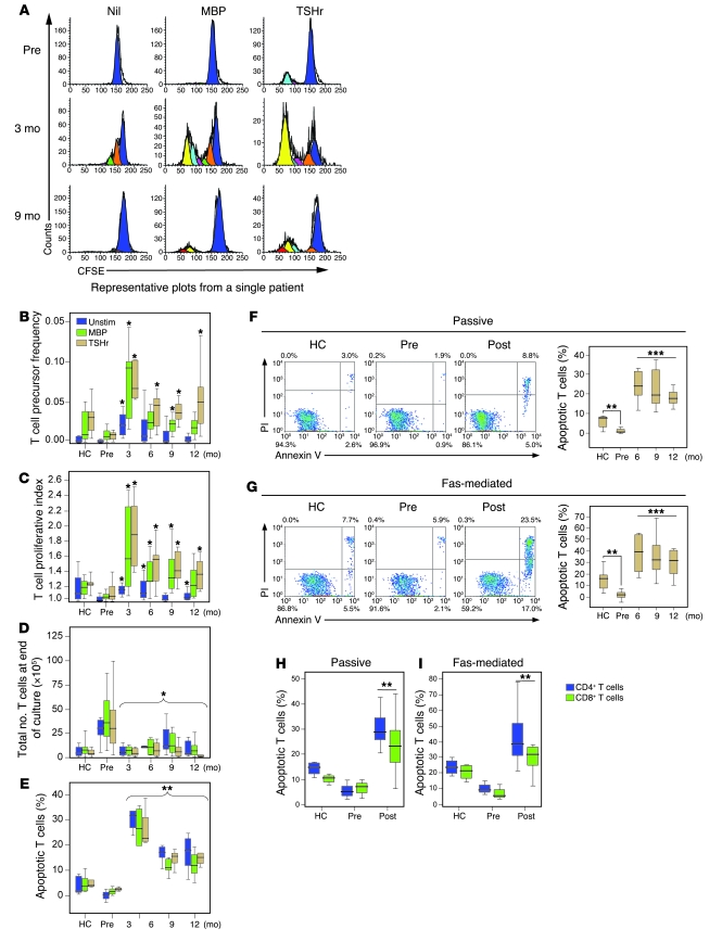
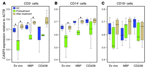
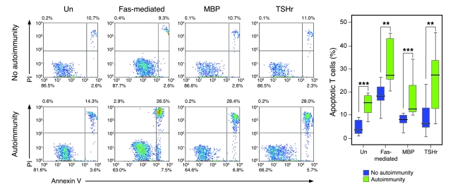
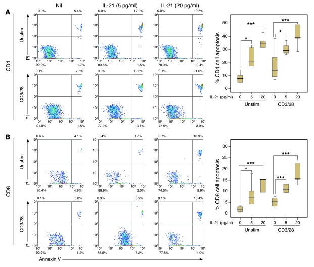
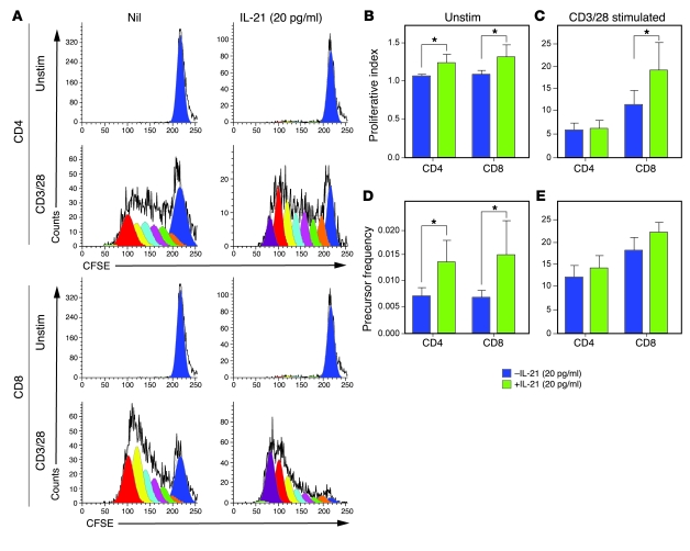
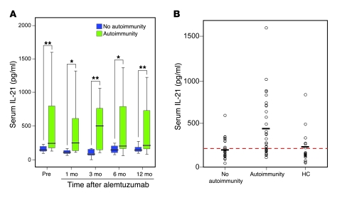
Phase II clinical trials revealed that the lymphocyte-depleting humanized monoclonal antibody alemtuzumab (Campath-1H) is highly effective in the treatment of early relapsing-remitting multiple sclerosis. However, 30% of patients develop autoimmunity months to years after pulsed exposure to alemtuzumab, usually targeting the thyroid gland and, more rarely, blood components. In this study, we show that autoimmunity arose in those patients with greater T cell apoptosis and cell cycling in response to alemtuzumab-induced lymphocyte depletion, a phenomenon that is driven by higher levels of IL-21. Before treatment, patients who went on to develop secondary autoimmunity had more than 2-fold greater levels of serum IL-21 than the nonautoimmune group. We suggest that serum IL-21 may, therefore, serve as a biomarker for the risk of developing autoimmunity months to years after alemtuzumab treatment. This has implications for counseling those patients with multiple sclerosis who are considering lymphocyte-depleting therapy with alemtuzumab. Finally, we demonstrate through genotyping that IL-21 expression is genetically predetermined. We propose that, by driving cycles of T cell expansion and apoptosis to excess, IL-21 increases the stochastic opportunities for T cells to encounter self antigen and, hence, for autoimmunity.


Figures

Figure 1
Figure 1. Sample and patient selection.
(A) Apoptosis and proliferation were performed on fresh ex vivo samples. Sixty-five patients were studied in total; of these, 29 were studied 9 months after alemtuzumab treatment. Of these, 10 patients met the study criteria for autoimmunity, and 10 patients met the criteria for no autoimmunity. The remaining 9 patients could not be categorized due to transient autoantibody production. (B) Serum IL-21 levels were determined in frozen pretreatment samples from 94 patients. Of these, 59 patients met the criteria for autoimmunity/no autoimmunity, of which, 30 patients had serial (3 monthly) samples available (15 with autoimmunity and 15 without autoimmunity). (C) Seventy-three patients gave consent for DNA analysis; of these, 27 patients met the criteria for autoimmunity and 23 patients met the study criteria for no autoimmunity.
Figure 2
Figure 2. A cross-sectional study of T cell proliferation and survival following alemtuzumab.
(A) Raw CFSE plots are shown, unstimulated (Nil) or following culture with MBP or TSHr. (B and C) Precursor frequency and proliferative index of T cells from healthy controls (HC), untreated patients (Pre), and patients 3, 6, 9, and 12 months after alemtuzumab, unstimulated or following culture with MBP or TSHr. (D) Total number of viable T cells after 10 days in culture. (E) Percentage of T cells apoptosing in response to no stimuli (Unstim) or following culture with MBP and TSHr. (F and G) Passive and Fas-mediated T cell apoptosis from healthy controls and patients before and after alemtuzumab (post) (right panels). Representative plots are shown (left panels). (H and I) Passive (Un) and Fas-mediated CD4+ and CD8+ T cell apoptosis from healthy controls, pretreatment patients, and at 9 months after alemtuzumab (*P < 0.05, **P < 0.01, ***P < 0.001). Box-and-whisker plots used depict the smallest value, lower quartile, median, upper quartile, and largest value. PI, propidium iodide.
Figure 3
Figure 3. Caspase-3 expression pretreatment and 9 months after alemtuzumab.
CASP3 mRNA expression relative to ACTB mRNA expression in (A) CD3+ T cells, (B) CD14+ monocytes, and (C) CD19+ B cells, either immediately ex vivo or following stimulation with MBP or polyclonal stimulation (anti-CD3/28 antibodies) (*P < 0.05). Box-and-whisker plots used depict the smallest value, lower quartile, median, upper quartile, and largest value.
Figure 4
Figure 4. Autoimmunity after alemtuzumab is associated with excessive T cell apoptosis.
Percentage of T cell apoptosis 9 months after alemtuzumab: passive, Fas-mediated, and in response to MBP and TSHr stimulation in those without autoimmunity (n = 10) and those with secondary autoimmunity (n = 10) (**P < 0.01, ***P < 0.001). Representative plots are shown. Box-and-whisker plots used depict the smallest value, lower quartile, median, upper quartile, and largest value.
Figure 5
Figure 5. rhIL-21 induces T cell apoptosis in vitro.
(A) CD4+ T cells and (B) CD8+ T cells, unstimulated or polyclonally stimulated (anti-CD3/CD28), apoptose in response to rhIL-21 in a dose-dependent manner. (*P < 0.05, ***P < 0.001). Representative plots are shown (left panels). Box-and-whisker plots used depict the smallest value, lower quartile, median, upper quartile, and largest value.
Figure 6
Figure 6. rhIL-21 induces T cell proliferation in vitro.
(A) Representative CFSE plots showing unstimulated and polyclonally stimulated (CD3/28) CD4+ T cells and CD8+ T cells with and without rhIL-21. (B and C) The proliferative index of unstimulated and polyclonally stimulated (anti-CD3/CD28) CD4+ and CD8+ T cells in response to rhIL-21. (D and E) Precursor frequency of unstimulated and polyclonally stimulated CD4+ and CD8+ T cells in response to rhIL-21 (*P < 0.05). Errors bars indicate 1 SD.
Figure 7
Figure 7. Serum IL-21.
(A) Serial serum IL-21, prior to, and after alemtuzumab treatment, in 15 patients with autoimmunity and in 15 patients without secondary autoimmunity. (B) Pretreatment serum IL-21 levels in the nonautoimmune (n = 27) and autoimmune (n = 32) cohorts compared with healthy controls (n = 19). The red dotted line represents serum IL-21 level of 210 pg/ml and the black horizontal lines represent mean values for each group (nonautoimmune, 206 pg/ml; autoimmune, 430 pg/ml; and healthy controls, 213 pg/ml) (*P < 0.05, **P < 0.01). Box-and-whisker plots used depict the smallest value, lower quartile, median, upper quartile, and largest value.

Comment in

References

    1. Coles A., et al. The window of therapeutic opportunity in multiple sclerosis. J. Neurol. 2006;253:98–108. doi: 10.1007/s00415-005-0934-5. - DOI - PubMed
    1. Coles A.J., et al. Alemtuzumab vs. interferon beta-1a in early multiple sclerosis. N. Engl. J. Med. 2008;359:1786–1801. doi: 10.1056/NEJMoa0802670. - DOI - PubMed
    1. Coles A.J., et al. Pulsed monoclonal antibody treatment and autoimmune thyroid disease in multiple sclerosis. Lancet. 1999;354:1691–1695. doi: 10.1016/S0140-6736(99)02429-0. - DOI - PubMed
    1. Chen F., et al. Characteristics of autoimmune thyroid disease occurring as a late complication of immune reconstitution in patients with advanced human immunodeficiency virus (HIV) disease. Medicine (Baltimore). 2005;84:98–106. doi: 10.1097/01.md.0000159082.45703.90. - DOI - PubMed
    1. Daikeler T., Tyndall A. Autoimmunity following haematopoietic stem-cell transplantation. Best. Pract. Res. Clin. Haematol. 2007;20:349–360. doi: 10.1016/j.beha.2006.09.008. - DOI - PubMed

Publication types

MeSH terms