. 2009 Sep;89(9):983-93.
doi: 10.1038/labinvest.2009.59. Epub 2009 Jun 22.

Slow progressive conduction and contraction defects in loss of Nkx2-5 mice after cardiomyocyte terminal differentiation

Affiliations

Slow progressive conduction and contraction defects in loss of Nkx2-5 mice after cardiomyocyte terminal differentiation

Morihiko Takeda et al. Lab Invest. 2009 Sep.

Abstract

Mutations in homeoprotein NKX2-5 are linked to human congenital heart disease, resulting in various cardiac anomalies, as well as in postnatal progressive conduction defects and occasional left ventricular dysfunction; yet the function of Nkx2-5 in the postnatal period is largely unexplored. In the heart, the majority of cardiomyocytes are believed to complete cell-cycle withdrawal shortly after birth, which is generally accompanied by a re-organization of chromatin structure shown in other tissues. We reasoned that the effects of the loss of Nkx2-5 in mice may be different after cell-cycle withdrawal compared with those of the perinatal loss of Nkx2-5, which results in rapid conduction and contraction defects within 4 days after the deletion of Nkx2-5 alleles (Circ Res. 2008;103:580). In this study, floxed-Nkx2-5 alleles were deleted using tamoxifen-inducible Cre transgene (Cre-ER) beginning at 2 weeks of age. The loss of Nkx2-5 beginning at 2 weeks of age resulted in conduction and contraction defects similar to the perinatal loss of Nkx2-5, however, with a substantially slower disease progression shown by 1 degrees atrioventricular block at 6 weeks of age (4 weeks after tamoxifen injections) and heart enlargement after 12 weeks of age (10 weeks after tamoxifen injections). The phenotypes were accompanied by a slower and smaller degree of reduction of several critical Nkx2-5 downstream targets that were observed in mice with a perinatal loss of Nkx2-5. These results suggest that Nkx2-5 is necessary for proper conduction and contraction after 2 weeks of age, but with a substantially distinct level of necessity at 2 weeks of age compared with that in the perinatal period.


Conflict of interest statement

Conflict of Interest:

None declared.

Figures

Figure 1
Figure 1. Time course study of tamoxifen-inducible targeting of Nkx2–5 beginning at 2 weeks of age
(A) Experimental time-course of Nkx2–5 knockout beginning at 2 weeks of age. (B) Northern blotting demonstrated that nearly complete loss of Nkx2–5 mRNA from 2.5 weeks (immediate after completion of four consecutive tamoxifen injections) to 3.5 weeks in flox/flox/Cre compared to flox/flox mice. (C) Western blotting demonstrated marked reduction of Nkx2–5 proteins (arrowheads) from 2.5 to 3.5 weeks of age in flox/flox/Cre mice. Non-specific band was detected at 2.5 weeks of age (marked with*). (D) Northern blotting demonstrated reduced expression of Nkx2–5 in flox/flox/Cre compared to flox/flox mice at 3.5 and 24 weeks of age after tamoxifen injections. fl, flox; wks, weeks.
Figure 2
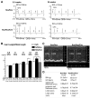
Figure 2. AV block and heart enlargement in Nkx2–5 knockout mice
(A) Examples of telemetry ECG recording at 3.5 and 24 weeks of age. At 24 weeks of age, telemetry ECG recording from flox/flox mouse showed normal sinus rhythm (left panel), however from flox/flox/Cre showed 2° AV block (right panel). See Table 1. (B) Heart weight/tibial length in Nkx2–5 knockout mice compared to control mice (flox/flox or flox/wild/Cre), with significant increase in heart weight/tibial length in Nkx2–5 knockout mice at 12 and 24 weeks of age. (C, D) Representative imaging of M-mode ultrasound microscope and echocardiographic indices of control and Nkx2–5 knockout mice approximately 24 weeks of age (mean ± S.E.) are shown. SCL, sinus cycle length (beta-to beat heart rate); BW, body weight; HR, heart rate; LVDd, left ventricular diastolic dimension; LVDs, left ventricular systolic dimension; %FS, percentage of left ventricular fractional shortening.
Figure 3
Figure 3. Nkx2–5 protein expression in contractile myocardium and AV node
(A) Co-immunostaining of Nkx2–5 and sarcomeric actinin demonstrate reduced expression of Nkx2–5 protein in ventricles dissected from Nkx2–5 mice at 3.5 weeks of age (left panels). Heart sections from 24 weeks old mice stained with Masson’s trichrome show no apparent interstitial fibrosis in Nkx2–5 knockout mice compared to control mice (right panels). Bars = 100 μm. (B) Tissue sections of 3.5 week old mouse heart demonstrates that AV node is positive for acetylcholine esterase staining (AchE) both in flox/flox and flox/flox/Cre mice. The adjacent tissue sections including AV node stained positive with anti-Nkx2–5 antibody (green) in flox/flox mice but was negative in flox/flox/Cre mice. Sarcomeric actinin staining is shown in red. Bars = 100 μm. (C) Whole mount acetylcholine esterase staining demonstrates AV node (brown staining, arrowheads) both in flox/flox and flox/flox/Cre mice at 3.5 and 24 weeks of age. Scale bars = 1 mm. (D) Surface area positive for acetylcholine esterase compared between flox/flox and flox/flox/Cre mice at 3.5 and 24 weeks of age (mean ± S.E.). *p < 0.05. (E) No apparent fibrosis was observed in Nkx2–5 knockout AV node analyzed by Masson’s trichrome staining at 24 weeks of age. AV nodal area is marked with arrows. Bars in low magnifications = 200 μm. Bars in high magnifications = 50 μm. (F) Nuclear density (number of nuclei/mm2) in flox/flox and flox/flox/Cre mice at 24 weeks of age. Total nuclear number counted was from a total of 3 different flox/flox or flox/flox/Cre mice. (G) HE-stained longitudinal sections of cardiomyocytes in AV node (left upper panels) and LV upper free wall (left bottom panels) were utilized for measurement of cell width at the nuclei level at 3.5 and 24 weeks of age. Bars = 20 μm. (H) AV nodal cell width (left panel) and LV cell width (right panel) compared between flox/flox and flox/flox/Cre mice at 3.5 and 24 weeks of age (mean ± S.E.). *p < 0.05. Total cell number counted was from a total of 3 different flox/flox or flox/flox/Cre mice at 3.5 weeks and 4 different flox/flox or flox/flox/Cre mice at 24 weeks. MV, mitral valve; Ao, aorta; IVS, interventricular septum.
Figure 4
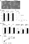
Figure 4. Increased cell size and Ca2+ handling defects in cardiomyocytes isolated from Nkx2–5 knockout heart
(A) Representative images of cardiomyocytes isolated from flox/flox (left) and flox/flox/Cre mice (right) at 24 weeks of age. Increased cell size was demonstrated in flox/flox/Cre mice. Bars = 100 μm. (B) Cell area (μm2), short axis (μm) and long axis (μm) of cardiomyocytes isolated from flox/flox or flox/flox/Cre mice (n=300 from 3 mice). (C) Distribution of the cell area of cardiomyocytes isolated from flox/flox and flox/flox/Cre mice. (D) Measurements of cardiac contraction and simultaneous Ca2+ transients in isolated cardiomyocytes from control flox/flox (white bars) or Nkx2–5 knockout (flox/flox/Cre) mice (black bars). Summarized data (mean ± S.E.) demonstrate that Nkx2–5 knockout cardiomyocytes show decreased Ca2+ amplitude, systolic fluorescence ratio, but preserved %fractional shortening, +dL/dT, −dL/dT and diastolic fluorescence ratio. *p < 0.05. (E) Increase of Ca2+ concentration in superfusate from 1.2 to 2.5 mM, increased %fractional shortening both in control and Nkx2–5 knockout cardiomyocytes, but was less in Nkx2–5 knockout cardiomyocytes compared to control cardiomyocytes.
Figure 5
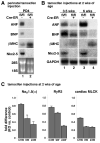
Figure 5. Expression of Nkx2–5 target genes in Nkx2–5 knockout hearts
(A) Northern blotting shows reduction of ANF and BNP mRNA and induction of βMHC mRNA in perinatal Nkx2–5 knockout hearts at PD4. (B) Northern blotting shows reduction of ANF and BNP mRNA at 3.5 and 6 weeks of age and induction of βMHC mRNA at 6 weeks of age in Nkx2–5 knockout hearts when tamoxifen was injected from 2 weeks of age. (C) Real-time RT-PCR shows fold difference (Nkx2–5 knockout vs. control) of mRNA of cardiac MLCK, Nav1.5(α) and RyR2 at 3.5 weeks, 9 weeks and 6 month of age. MLCK, myosin light chain kinase; RyR2, ryanodine receptor 2; KO, knockout.

Similar articles

Cited by

References

    1. Harvey RP. NK-2 homeobox genes and heart development. Dev Biol. 1996;178(2):203–216. - PubMed
    1. Harvey RP, Rosenthal N. Heart Development. Academic Press; 1999.
    1. Lints TJ, Parsons LM, Hartley L, Lyons I, Harvey RP. Nkx-2.5: a novel murine homeobox gene expressed in early heart progenitor cells and their myogenic descendants. Development. 1993;119(2):419–431. - PubMed
    1. Komuro I, Izumo S. Csx: a murine homeobox-containing gene specifically expressed in the developing heart. Proc Natl Acad Sci U S A. 1993;90(17):8145–8149. - PMC - PubMed
    1. Kasahara H, Bartunkova S, Schinke M, Tanaka M, Izumo S. Cardiac and extracardiac expression of Csx/Nkx2.5 homeodomain protein. Circ Res. 1998;82(9):936–946. - PubMed

Publication types

MeSH terms