. 2009 Sep;89(9):1007-17.
doi: 10.1038/labinvest.2009.58. Epub 2009 Jun 22.

Osteopetrosis with micro-lacunar resorption because of defective integrin organization

Affiliations

Osteopetrosis with micro-lacunar resorption because of defective integrin organization

Harry C Blair et al. Lab Invest. 2009 Sep.

Abstract

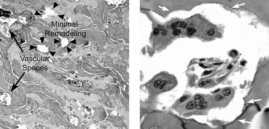
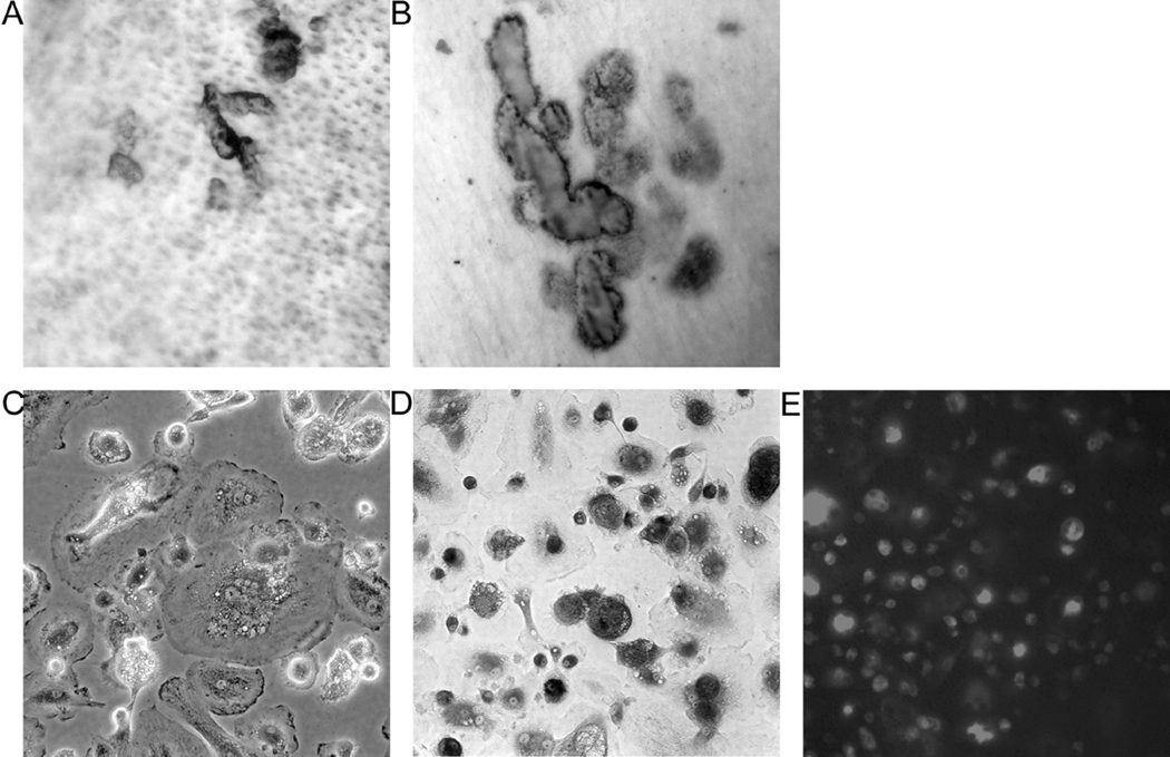
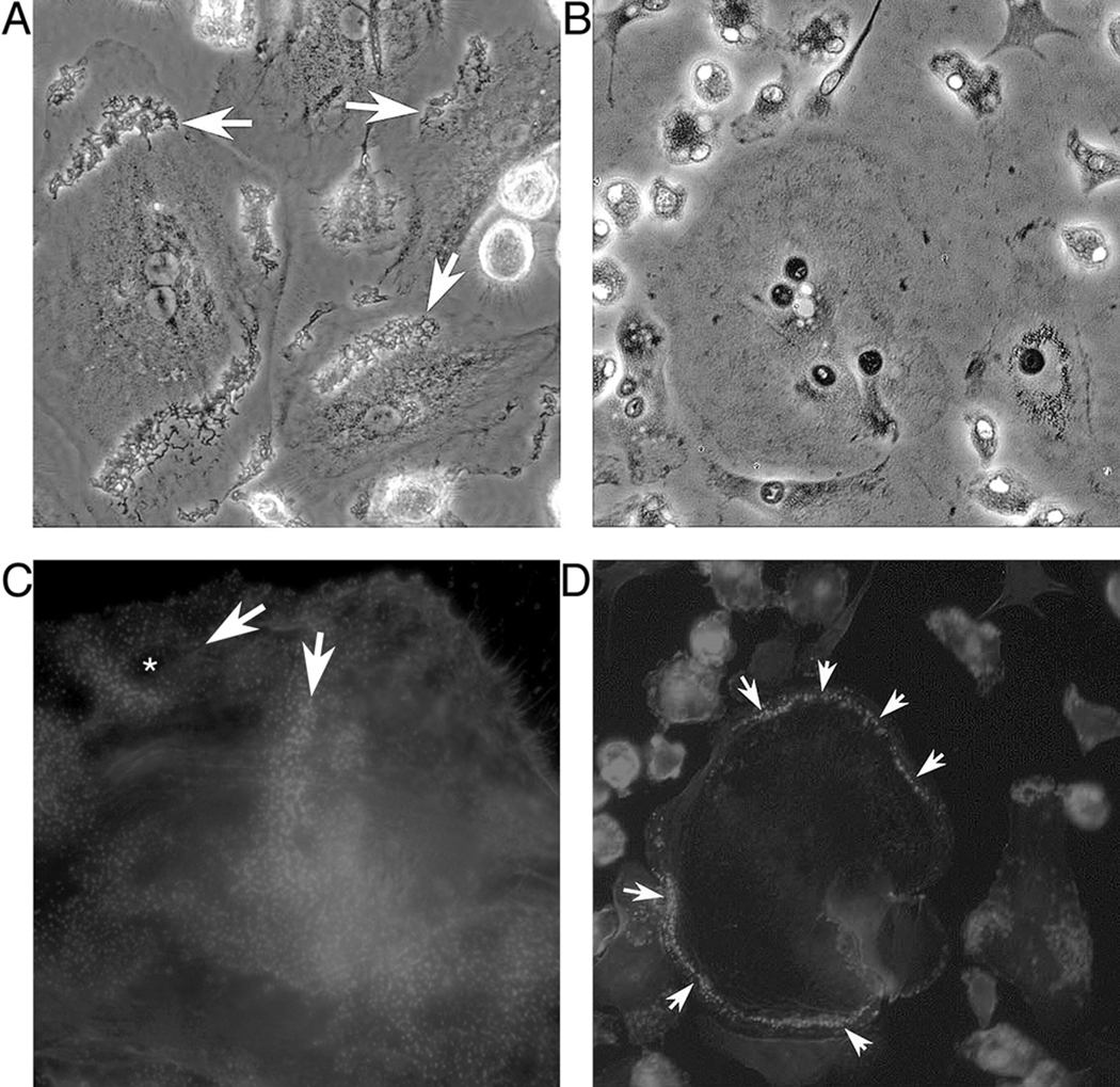
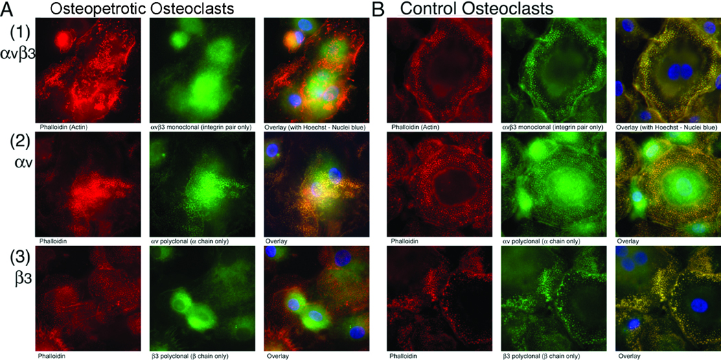
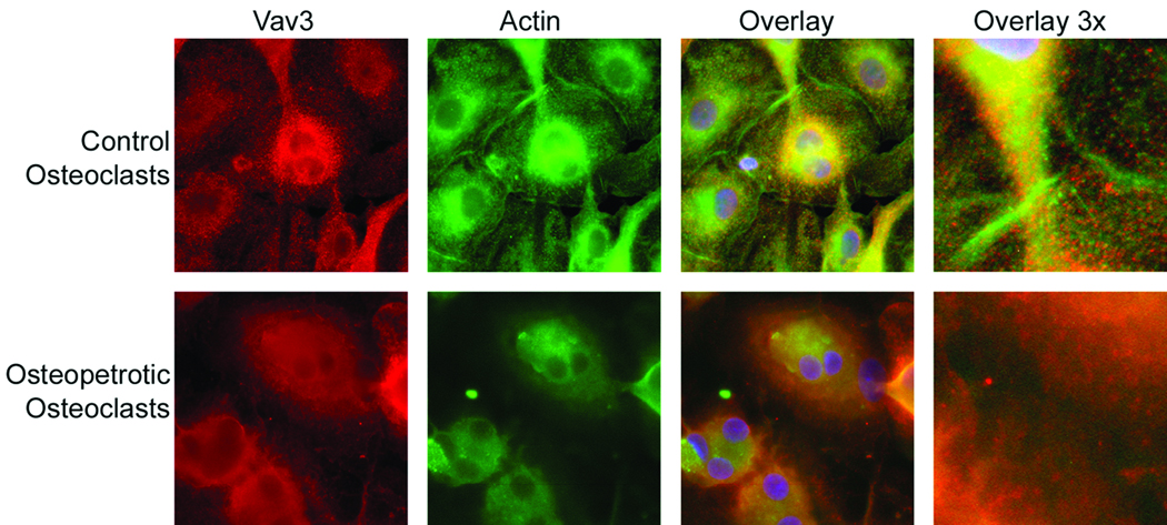
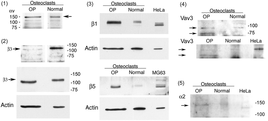
In vitro differentiated monocytes were used to characterize the cellular defect in a type of osteopetrosis with minimally functional osteoclasts, in which defects associated with common causes of osteopetrosis were excluded by gene sequencing. Monocytes from the blood of a 28-year-old patient were differentiated in media with RANKL and CSF-1. Cell fusion, acid compartments within cells, and tartrate resistant acid phosphatase (TRAP) activity were normal. However, the osteoclasts made abnormally small pits on the dentine. Phalloidin labeling showed that the cell attachments lacked the peripheral ring structure that supports lacunar resorption. Instead, the osteoclasts had clusters of podosomes near the center of cell attachments. Antibody to the alphavbeta3 integrin pair or to the C-terminal of beta3 did not label podosomes, but antibody to alphav labeled them. Western blots using antibody to the N-terminal of beta3 showed a protein of reduced size. Integrins beta1 and beta5 were upregulated, but, in contrast to observations in beta3 defects, alpha2 had not increased. The rho-GTP exchange protein Vav3, a key attachment organizing protein, did not localize normally with peripheral attachment structures. Vav3 forms of 70 kD and 90 kD were identified on western blots. However, the proteins beta3 integrin, Vav3, Plekhm1, and Src, implicated in attachment defects, had normal exon sequences. In this new type of osteopetrosis, the integrin-organizing complex is dysfunctional, and at least two attachment proteins may be partially degraded.


Figures

Figure 1
Figure 1. Features of bone from a biopsy of the iliac crest
A. Severe osteopetrosis with minimal remodeling including some vascular spaces. Although the subject survived for 28 years prior to transplant, the major part of the bone was retained cartilage (lightly stained acellular material) with the balance mainly composed of primary spongiosa, and a few spaces. Low power (10× objective); the field shown is 800 µm across. B. Osteoclasts with shallow resorptive pits. Osteoclasts were present and appeared normal by light microscopy, although pits were small and shallow relative to the number of multinucleated cells present. High power (40× objective); the field shown is 110 µm across.
Figure 2
Figure 2. Osteoclasts formed in vitro from osteopetrotic monocytes make small pits but have normal multinucleation and tartrate resistant acid phosphatase expression
A–B Pit assays from osteopetrotic and control osteoclasts. Osteoclasts from the osteopetrotic patient produced pits (A), but the pits averaged 10–15 µm in diameter, much smaller than the osteoclasts themselves (Figure 1 and C–D below). Control osteoclasts produced pits averaging 40 µm in diameter (B). Fields 450 µm across (20× objective) are shown. Cells produced in vitro were incubated on whale dentine for 12 days. The substrate was cleaned by sonication and labeled with toluidine blue to show partially degraded matrix proteins at pit sites. C. Osteopetrotic osteoclasts in vitro. Phase microscopy showed large multinucleated cells, typical of RANKL stimulated cell differentiation in vitro. The field shown is 280 µm across. D. tartrate-resistant acid phosphatase activity of osteopetrotic osteoclasts formed in vitro. Tartrate resistant acid phosphatase activity was completely typical of osteoclasts in vitro. This frame a bright-field image 450 µm across, the same magnification as A and B. Note that the pits in B have similar sizes to the large rounded cells expressing large amounts of TRAP, but that the pits in A are much smaller (see also Figure 1B). E. Lysotracker red DND labeling. Cells on glass contain abundant acidic vacuoles, the same as in normal osteoclasts (not illustrated) [4]. The field is 450 µm across.
Figure 3
Figure 3. High resolution phase and phalloidin rhodamine labeling show podosomal anomalies in osteopetrotic osteoclasts
Cells were grown on glass cover slips. The fields shown were taken with 40× objectives and all photographs are 220 µm across. A, C. Osteoclasts from the osteopetrotic patient. In phase contrast at high power (A) prominent cell membrane structures often occurred in broad regions of the cells (arrows). Phalloidin rhodamine labeling (C) showed very large clusters and bands of podosomes sometimes in peripheral bands and in some cases across the center of the cell attachment (arrows). These ringed some spaces (asterisk) but typically these were much smaller than the cell diameters, and highly variable. A and C are photographs of two similar fields. B, D. Control normal osteoclasts showing peripheral cell attachment structure. In contrast to osteopetrotic cells, normal osteoclasts have well defined borders in phase (B) that correspond to one or more regular and well defined peripheral rings of podosomes (Arrowheads, D). The cells shown in B and D are the same field.
Figure 4
Figure 4. Integrin distribution in control and osteopetrotic osteoclasts
Cells grown as in Figures 1–2 were labeled for actin (phalloidin, red), antibodies to integrins specified (green), and Hoechst dye (nuclei, blue). Fields are each 85 µm across. A. Podosomes from osteoclasts of the affected subject label poorly for αvβ3 integrin or β3 subunits, but include αv integrin. The left column (red) shows actin with prominent podosomes in abnormal distribution (larger fields shown in Figure 3). Monoclonal antibody for the αvβ3 pair (1, top row) and polyclonal antibodies to αv (2, middle row) and β3 N-terminal (3, bottom row) were used to label integrins (second column, green). There was essentially no colocalization αvβ3 with podosomes (third column, yellow shows colocalization). Labeling with anti-N-terminal β3 was weak and did not colocalize with actin. This may reflect degradation of the β3 C-terminal (see Figure 6). There was colocalization of αv with podosomes (third column from left), although podosomes were abnormally distributed as in Figure 3. B. Podosomes from normal osteoclasts label for αvβ3 integrin, and for αv or β3 integrin subunits. The photographs parallel those of the osteopetrotic cells (A). The osteoclasts have prominent peripheral rings of podosomes that label well with the integrin pair and with each integrin subunit (yellow in merged images, right column). No labeling of control or osteopetrotic cells for α2 integrin was detected in similar assays (not shown; α2 integrin is not expressed in major amounts in these cells, see Figure 6).
Figure 5
Figure 5. Dysfunctional distribution of Vav3 in osteopetrotic cells with abnormal attachment
Vav3 is critical to the organization of the cellular attachment and membrane receptors. Vav3 is associated with attachment structures in the periphery of control cells (top row). Vav3 was not associated with actin or podosomes in the periphery of osteopetrotic cells (bottom row). The relationship of Vav3, around the periphery of podosomes and near actin filaments, is seen most clearly at very high magnification power (right frames, 30 µm across). Other frames are 85 µm square.
Figure 6
Figure 6. Western blots for integrin subunits and Vav3 in osteopetrotic osteoclasts and control cells
Blots used 10 µg of cell lysate protein separated on 8 or 10% SDS-PAGE and polyclonal antibodies visualized by enhanced chemiluminescence (see Methods). (1) Control and osteopetrotic cells showed similar results with antibody to the αv integrin subunit. (2) Re-blot of the membrane (1) for the C terminal region of the β3 subunit was very weak in the osteopetrotic patient. A second blot using an antibody to the β3 subunit N terminal region was showed similar β3 in control and osteopetrotic cell (OP) cell lysates, although the β3 is probably differently processed since its nucleotide sequence was normal (see text). Actin re-blot is shown as a protein loading control in the second blot. Under the conditions used, actin is near the front and OP lysates, which contained more salt than controls, expanded toward the front. (3) Osteoclasts with β3 defects have compensatory increases in β1 or β5. Additional blots using HeLa cells as a positive control for β1 and MG63 cells as a control for β5, since these integrin chains are expressed at low levels in normal osteoclasts. Both β1 and β5 were present in higher amounts in osteopetrotic cells than in normal controls. (4) Re-blots of membranes from (1) and (3) for Vav3. The OP patient showed two isoforms, one ~20 kD smaller than the normal form, which is also abundant in the HeLa cells. (5) The α2 integrin subunit is a minor protein, barely detectable, in normal or OP osteoclasts. Re-blot of the membrane in (3) upper panel. This is in keeping with normal αv expression, but contrasts with findings in patients with isolated β3 defects (see text). Re-blot of (3).

References

    1. Tolar J, Teitelbaum SL, Orchard PJ. Osteopetrosis. N Engl J Med. 2004;351:2839–2349. - PubMed
    1. Blair HC, Teitelbaum SL, Ghiselli R, et al. Osteoclastic bone resorption by a polarized vacuolar proton pump. Science. 1989;245:855–857. - PubMed
    1. Li YP, Chen W, Liang Y, et al. Atp6i-deficient mice exhibit severe osteopetrosis due to loss of osteoclast-mediated extracellular acidification. Nat Genet. 1999;23:447–451. - PubMed
    1. Blair HC, Borysenko CW, Villa A, et al. In vitro differentiation of CD14 cells from osteopetrotic subjects: contrasting phenotypes with TCIRG1, CLCN7, and attachment defects. J Bone Miner Res. 2004;19:1329–1338. - PubMed
    1. Scheel O, Zdebik AA, Lourdel S, et al. Voltage-dependent electrogenic chloride/proton exchange by endosomal CLC proteins. Nature. 2005;436:424–427. - PubMed

Publication types

MeSH terms