. 2009 Jun 23;4(6):e6020.
doi: 10.1371/journal.pone.0006020.

Chlamydia pneumoniae hides inside apoptotic neutrophils to silently infect and propagate in macrophages

Affiliations

Chlamydia pneumoniae hides inside apoptotic neutrophils to silently infect and propagate in macrophages

Jan Rupp et al. PLoS One. .

Abstract

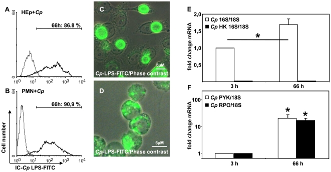
Background: Intracellular pathogens have developed elaborate strategies for silent infection of preferred host cells. Chlamydia pneumoniae is a common pathogen in acute infections of the respiratory tract (e.g. pneumonia) and associated with chronic lung sequelae in adults and children. Within the lung, alveolar macrophages and polymorph nuclear neutrophils (PMN) are the first line of defense against bacteria, but also preferred host phagocytes of chlamydiae.

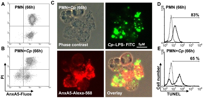
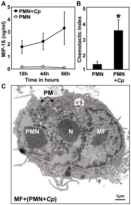
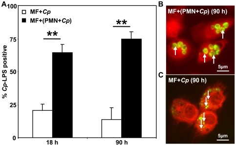
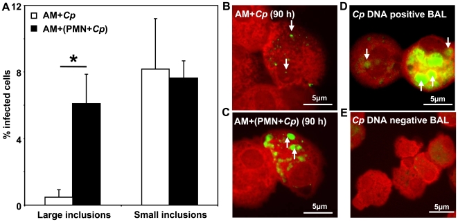
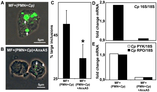
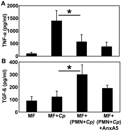
Methodology/principal findings: We could show that C. pneumoniae easily infect and hide inside neutrophil granulocytes until these cells become apoptotic and are subsequently taken up by macrophages. C. pneumoniae infection of macrophages via apoptotic PMN results in enhanced replicative activity of chlamydiae when compared to direct infection of macrophages, which results in persistence of the pathogen. Inhibition of the apoptotic recognition of C. pneumoniae infected PMN using PS- masking Annexin A5 significantly lowered the transmission of chlamydial infection to macrophages. Transfer of apoptotic C. pneumoniae infected PMN to macrophages resulted in an increased TGF-ss production, whereas direct infection of macrophages with chlamydiae was characterized by an enhanced TNF-alpha response.

Conclusions/significance: Taken together, our data suggest that C. pneumoniae uses neutrophil granulocytes to be silently taken up by long-lived macrophages, which allows for efficient propagation and immune protection within the human host.


Conflict of interest statement

Competing Interests: The authors have declared that no competing interests exist.

Figures

Figure 1
Figure 1. C. pneumoniae survives inside PMN.
C. pneumoniae (Cp) infected HEp-2 cells and neutrophils (PMN) were analyzed 66 h p.i. for intracellular positivity of chlamydial LPS by FACS analysis (A, B; representative experiment out of 3) and fluorescence microscopy (C, D; magnification 630×). To analyze intracellular progeny of C. pneumoniae in PMN we performed real-time RT-PCRs of the 16S rRNA in comparison to host cell 18S rRNA using the ΔΔct- method for relative quantification. Viable (open bars) but not heat-killed (HK, closed bars) chlamydiae showed a significant increase in 16S rRNA expression (E) within 66 h p.i. (p = 0.02; n = 3). Enhanced transcriptional activity of C. pneumoniae inside PMN was proven by determining the expression of chlamydial genes pyk (open bars, p = 0.03) and rpoA (closed bars, p = 0.01) mRNA (F; n = 3).
Figure 2
Figure 2. C. pneumoniae hides inside PS- positive PMN.
To analyze early and late apoptotic markers, PMN were infected with C. pneumoniae or left in medium alone. Using Anxa5-fluos and propidium iodide (PI) staining we could show that both non- infected (>95%, A) and C. pneumoniae- infected (82%±12%, B) PMN become PS- positive within 66 h without significant changes in the amount of PI- positive necrotic cells (representative experiment out of 3). To visualize whether C. pneumoniae- infected PMN stain positive for PS we performed a double staining using a FITC- labeled anti-C. pneumoniae LPS mAb and Alexa568-labeled AnxA5 (C). In contrast, late apoptotic cells as determined by TUNEL staining (closed line) were more often found in the medium control than in C. pneumoniae- infected PMN (D, E; representative experiment out of 3).
Figure 3
Figure 3. Increased MIP-1ß release in C. pneumoniae infected PMN.
PMN were infected with C. pneumoniae (closed circles) or mock infected with HEp-2 lysates (open circles) (A). Increased secretion of MIP-1ß was observed in the supernatants of C. pneumoniae- infected PMN at the given time points by ELISA (A). The chemotactic index (specific migration/migration towards medium) indicates that monocytes are significantly better attracted by supernatants taken from C. pneumoniae- infected PMN than mock- infected control cells (B, p = 0.04; n = 3). Using transmission electron microscopy we could show that C. pneumoniae- infected PMN are engulfed by monocyte- derived macrophages (C). Arrows indicate the phagosomal membrane (PM) of the macrophage; containing a complete apoptotic PMN with condensed nucleus (N) (bar equals 1 µm, magnification 6000×).
Figure 4
Figure 4. PMN passage increases chlamydial transmission to macrophages.
C. pneumoniae alone (open bars) or C. pneumoniae- infected PMN (closed bars, 66 h p.i.) were co incubated with autologous macrophages (MF) at ratio of 1∶1 (A–C). The amount of C. pneumoniae- positive (C. pneumoniae LPS- staining) MF increased significantly after 18 and 90 h when MF were co incubated with C. pneumoniae- infected apoptotic PMN (A, p = 0.004), but not upon direct C. pneumoniae infection. Fluorescence microscopy revealed that co incubation of MF with C. pneumoniae- infected apoptotic PMN for 90 h resulted in multiple large inclusions (B) whereas directly infected MF showed smaller “persistent-like” inclusions (C; magnification 630×).
Figure 5
Figure 5. Alveolar macrophages harbor intracellular chlamydiae in vivo.
Alveolar macrophages (AM) isolated from BAL fluids of C. pneumoniae- DNA negative patients were either directly infected with C. pneumoniae (open bars) or co incubated with C. pneumoniae infected PMN at a ratio of 1∶1 (closed bars, 66 h p.i.). The percentage of AM harvesting large and small inclusions were calculated 90 h p.i. by counting a minimum of 200 cells/slide using a FITC- labeled anti- C. pneumoniae LPS- staining protocol (A). Significantly more large chlamydial inclusions were detected in AM co incubated with C. pneumoniae- infected apoptotic PMN than in directly infected AM (A–C, p = 0.02). In addition, we analyzed AM from BAL fluids of patients suffering from community-acquired pneumonia (CAP) by fluorescence microscopy (D, E; representative experiment out of 4), showing both small, persistent-like inclusions (downward arrows) and large inclusions (upward arrows) in C. pneumoniae- DNA positive BAL (D) but not in C. pneumoniae- DNA negative BAL (E).
Figure 6
Figure 6. PS- dependent transmission of C. pneumoniae infection.
Blocking of phosphatidylserine (PS) expression on C. pneumoniae- infected PMN by preincubation with recombinant AnxA5 significantly reduced the uptake of chlamydiae (A, B) and the formation of large inclusions (C, p = 0.02; n = 3) as shown by fluorescence microscopy with FITC- labeled anti- C. pneumoniae LPS- staining (representative experiment out of 3, magnification 630×) and calculation of a minimum of 200 cells/slide (C). Replicative activity of C. pneumoniae, indicated by the amount of 16S rRNA (D), pyk (open bars) and rpo (closed bars) mRNA expression compared to host cell 18S rRNA expression (E), decreased in MF when C. pneumoniae- infected PMN were preincubated with AnxA5 (representative experiment out of 3).
Figure 7
Figure 7. Silencing of MF immune response to chlamydial infection by PMN passage.
Immune responses of directly C. pneumoniae- infected MF were analyzed in comparison to MF either co incubated with C. pneumoniae- infected PMN (66 h p.i.) or with C. pneumoniae- infected PMN after precinubation with recombinant AnxA5. Within 18 h p.i. supernatants of MF were collected to determine total amounts of TNF-α (A) and TGF-ß (B) by ELISA. Direct infection of MF with C. pneumoniae significantly increased TNF-α production (A, p = 0.01; n = 3), whereas the uptake of C. pneumoniae- infected apoptotic PMN was characterized by a significant up regulation of TGF-ß in MF (B, p = 0.02; n = 3).

References

    1. Gieffers J, van Zandbergen G, Rupp J, Sayk F, Kruger S, Ehlers S, Solbach W, Maass M. Phagocytes transmit Chlamydia pneumoniae from the lungs to the vasculature. Eur Respir J. 2004;23:506–510. - PubMed
    1. Yang ZP, Cummings PK, Patton DL, Kuo CC. Ultrastructural lung pathology of experimental Chlamydia pneumoniae pneumonitis in mice. J Infect Dis. 1994;170:464–467. - PubMed
    1. Jahn HU, Krull M, Wuppermann FN, Klucken AC, Rosseau S, Seybold J, Hegemann JH, Jantos CA, Suttorp N. Infection and activation of airway epithelial cells by Chlamydia pneumoniae. J Infect Dis. 2000;182:1678–1687. - PubMed
    1. van Zandbergen G, Gieffers J, Kothe H, Rupp J, Bollinger A, Aga E, Klinger M, Brade H, Dalhoff K, Maass M, Solbach W, Laskay T. Chlamydia pneumoniae multiply in neutrophil granulocytes and delay their spontaneous apoptosis. J Immunol. 2004;172:1768–1776. - PubMed
    1. Bartels C, Maass M, Bein G, Malisius R, Brill N, Bechtel JF, Sayk F, Feller AC, Sievers HH. Detection of Chlamydia pneumoniae but not cytomegalovirus in occluded saphenous vein coronary artery bypass grafts. Circulation. 1999;99:879–882. - PubMed

Publication types