Review
. 2009 Oct;66(19):3137-47.
doi: 10.1007/s00018-009-0070-y. Epub 2009 Jun 23.

The current structural and functional understanding of APOBEC deaminases

Affiliations
Review

The current structural and functional understanding of APOBEC deaminases

Ronda Bransteitter et al. Cell Mol Life Sci. 2009 Oct.

Abstract

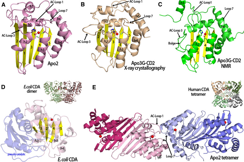
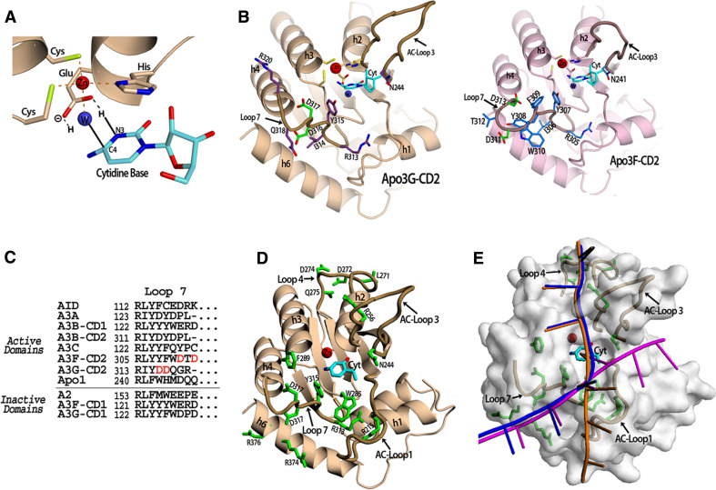
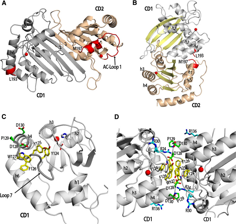
The apolipoprotein B mRNA-editing enzyme catalytic polypeptide (APOBEC) family of cytidine deaminases has emerged as an intensively studied field as a result of their important biological functions. These enzymes are involved in lipid metabolism, antibody diversification, and the inhibition of retrotransposons, retroviruses, and some DNA viruses. The APOBEC proteins function in these roles by deaminating single-stranded (ss) DNA or RNA. There are two high-resolution crystal structures available for the APOBEC family, Apo2 and the C-terminal catalytic domain (CD2) of Apo3G or Apo3G-CD2 [Holden et al. (Nature 456:121-124, 2008); Prochnow et al. (Nature 445:447-451, 2007)]. Additionally, the structure of Apo3G-CD2 has also been determined using NMR [Chen et al. (Nature 452:116-119, 2008); Furukawa et al. (EMBO J 28:440-451, 2009); Harjes et al. (J Mol Biol, 2009)]. A detailed structural analysis of the APOBEC proteins and a comparison to other zinc-coordinating deaminases can facilitate our understanding of how APOBEC proteins bind nucleic acids, recognize substrates, and form oligomers. Here, we review the recent development of structural and functional studies that apply to Apo3G as well as the APOBEC deaminase family.


Figures

Fig. 1
Fig. 1
Comparison of APOBEC and fntCDA structures. a An Apo2 monomer (PDB 2nyt, chain A-closed loop conformation) showing the catalytic residues and a zinc molecule (red sphere) in the active center. b The crystal structure of an Apo3G-CD2 monomer (PDB 3e1u) contains 5 beta strands similar to Apo2 and other deaminases. c An Apo3G-CD2 structure solved by NMR (PDB 2KBO) which is very similar to the other APOBEC structures (see a and b). In contrast to the Apo3G-CD2 crystal structure, the NMR structure has a discontinuous beta 2 strand that contains a bulge. Also, AC-Loop 1 and h1 are positioned differently in comparison to the Apo3G-CD2 structure in (b). d The structure of an ECDA monomer containing a pseudo-catalytic domain (light blue) connected to the catalytic domain (light purple with yellow beta strands) via a long flexible loop. The active center zinc is represented as a red sphere. Unlike the APOBEC structures that contain a long h4 and h6, the equivalent ECDA h4 forms a long flexible loop and small 310 helices that connect to the pseudo-catalytic domain (inset). The square-shaped E. coli CDA dimer (PDB 1ALN) with the monomers shown in green and purple, each containing an active center zinc (red sphere). e The Apo2 tetramer (PDB 2nyt) displays a novel type of oligomerization forming dimers through a head–head interaction. H4 and h6 allow for the extended tetramer formation with interactions occurring though residues from Loop 7, AC-Loop 1, h4 and h6. The closed AC-Loop 1 conformations found in the inner monomers (light pink and light blue) at the tetramer interface may block substrate access to these active centers. Each monomer is colored differently with a zinc ion in the active center (red sphere) (inset). The human CDA square-shaped tetramer (PDB 1mq0) with the monomers shown in different colors and the zinc ion represented as a red sphere
Fig. 2
Fig. 2
APOBEC DNA Binding. a The APOBEC active site containing a cytidine base. The Apo3G-CD2 crystal structure was superimposed with the mouse CDA co-crystal structure with cytidine in the active site (PDB 2fr6) in order to place the cytidine base in this position (shown in cyan). Three residues (2 cysteines and 1 histidine) coordinate the zinc ion (red sphere) along with a water molecule (blue sphere). The conserved glutamate shuffles the protons from the water molecule to the cytidine during the hydrolytic deamination. The proposed mechanism occurs via the glutamate acting to protonate N3 of the cytidine ring by transferring a hydrogen molecule from the water. Next, the activated water molecule (a Zn-hydroxide) attacks the C4 of the cytidine ring resulting in the release of ammonia. b The Apo3G-CD2 structure (PDB 3e1u) which shows the residues on Loop 7 which influence APOBEC substrate specificity. Residues previously shown to be important for Apo3G-CD2 substrate specificity (D316 and D317) are shown in green. Other residues on Loop 7 (shown in purple) may contact the residues neighboring the target cytidine through electrostatic, hydrophobic or base stacking interactions. N244 on AC-Loop 3 is conserved throughout the zinc-dependent deaminase family and has been shown to contact the target base directly (inset). The model of Apo3F-CD2 shows the predicted Loop 7 residues. The SWISS-MODEL homology modeling program was used to generate the model using the Apo3F sequence and the Apo3G-CD2 crystal structure (PDB 3e1u) as a template [106]. Residues D311 and D313 shown in green on Loop 7 have previously been shown to be important in substrate specificity. The conserved N241 which may directly contact the target cytidine is shown on AC-Loop 3. c An alignment of APOBEC proteins showing residues predicted to be in Loop 7. The domains are categorized according to whether they have been shown to be active for deamination activity. d Apo3G-CD2 residues previously reported to be important for DNA binding. The cytidine base is colored cyan and DNA binding residues are colored green. e The proposed Apo3G-CD2 DNA binding models. The target cytidine (cyan) is shown in its proposed position in the active center (see a). The DNA binding model proposed by Holden et al. [1] is shown in magenta. The model proposed by the Chen et al. and Furukawa et al. is shown in orange [3, 4]. A combination of both models is shown in blue
Fig. 3
Fig. 3
Models of Apo3G monomers and dimers. a A model of an Apo3G monomer whereby the two domains fold similar to an Apo2 dimer by pairing of the β2 strands. The inter domain connection between Apo3G-CD1 and Apo3G-CD2 would have to be made through an additional three amino acids (194–196) that could connect Apo3G-CD1 h6 (L193) to h1 of Apo3G-CD2 (M197) (connection to be made is highlighted in red). The SWISS-MODEL protein homology modeling program was used to generate the Apo3G-CD1 model using an Apo2 monomer in a closed conformation as a template [106]. PyMOL was used to generate the full-length Apo3G monomer using an Apo2 dimer as a template (PDB 2nyt): the Apo3G-CD1 model was aligned with one Apo2 monomer and the structure of Apo3G-CD2 (PDB 3e1u) with the other Apo2 monomer [107]. Apo3G-CD1 is colored grey, Apo3G-CD2 is colored wheat and the two zinc molecules are represented by red spheres. b A different model of an Apo3G monomer adapted from Harjes et al. where the two active center domains of Apo3G-CD1 and Apo3G-CD2 lie on opposite faces (zinc ion represented by red spheres). The Apo3G-CD2 structure (3e1u) was aligned with the Apo3G-CD1 model according to Harjes et al. [5]. c A model of Apo3G-CD1 displaying the residues on Loop 7 important for dimerization (yellow residues) and Vif binding (green residues). d A model of the proposed Apo3G dimer CD1–CD1 (head–head) interface based off the Apo2 tetramer interface (PDB 2nyt). Yellow colored residues (Y124, Y125 and W127) have been proposed to be important for dimerization and virion incorporation. The residues (D128, P129 and D130) that interact with Vif are colored green. Residues important for binding RNA, R24, R30 and R136, are colored cyan

References

    1. Holden LG, Prochnow C, Chang PY, Bransteitter R, Chelico L, Sen U, Stevens RC, Goodman MF, Chen XS. Crystal structure of the anti-viral APOBEC3G catalytic domain and functional implications. Nature. 2008;456:121–124. - PMC - PubMed
    1. Prochnow C, Bransteitter R, Klein MG, Goodman MF, Chen XS. The APOBEC-2 crystal structure and functional implications for the deaminase AID. Nature. 2007;445:447–451. - PubMed
    1. Chen KM, Harjes E, Gross PJ, Fahmy A, Lu Y, Shindo K, Harris RS, Matsuo H. Structure of the DNA deaminase domain of the HIV-1 restriction factor APOBEC3G. Nature. 2008;452:116–119. - PubMed
    1. Furukawa A, Nagata T, Matsugami A, Habu Y, Sugiyama R, Hayashi F, Kobayashi N, Yokoyama S, Takaku H, Katahira M. Structure, interaction and real-time monitoring of the enzymatic reaction of wild-type APOBEC3G. EMBO J. 2009;28:440–451. - PMC - PubMed
    1. Harjes E, Gross PJ, Chen KM, Lu Y, Shindo K, Nowarski R, Gross JD, Kotler M, Harris RS, Matsuo H (2009) An extended structure of the APOBEC3G catalytic domain suggests a unique holoenzyme model. J Mol Biol (in press) - PMC - PubMed

Publication types

LinkOut - more resources