. 2009 Aug;44(4):213-23.
doi: 10.1007/s10858-009-9333-z. Epub 2009 Jun 23.

TALOS+: a hybrid method for predicting protein backbone torsion angles from NMR chemical shifts

Affiliations

TALOS+: a hybrid method for predicting protein backbone torsion angles from NMR chemical shifts

Yang Shen et al. J Biomol NMR. 2009 Aug.

Abstract

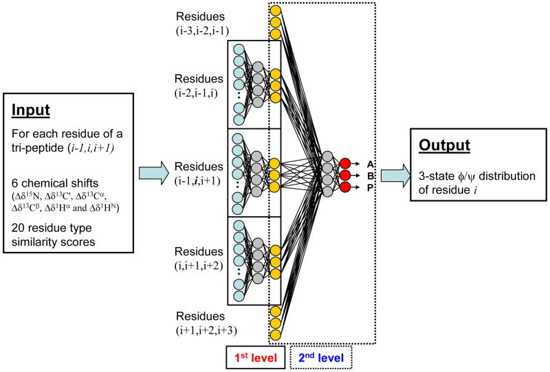
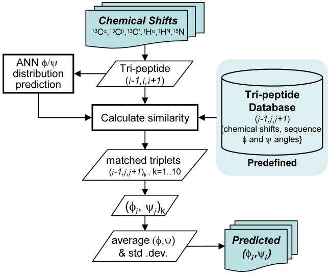
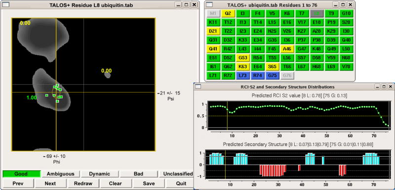
NMR chemical shifts in proteins depend strongly on local structure. The program TALOS establishes an empirical relation between 13C, 15N and 1H chemical shifts and backbone torsion angles phi and psi (Cornilescu et al. J Biomol NMR 13 289-302, 1999). Extension of the original 20-protein database to 200 proteins increased the fraction of residues for which backbone angles could be predicted from 65 to 74%, while reducing the error rate from 3 to 2.5%. Addition of a two-layer neural network filter to the database fragment selection process forms the basis for a new program, TALOS+, which further enhances the prediction rate to 88.5%, without increasing the error rate. Excluding the 2.5% of residues for which TALOS+ makes predictions that strongly differ from those observed in the crystalline state, the accuracy of predicted phi and psi angles, equals +/-13 degrees . Large discrepancies between predictions and crystal structures are primarily limited to loop regions, and for the few cases where multiple X-ray structures are available such residues are often found in different states in the different structures. The TALOS+ output includes predictions for individual residues with missing chemical shifts, and the neural network component of the program also predicts secondary structure with good accuracy.


Figures

Figure 1
Figure 1
Prediction of the three-state φ/ψ distribution using a neural network with a 3–3 ANN model. (A) φ/ψ distribution of the residues in the 200-protein TALOS database. Boxed areas marking the 3-state φ/ψ regions for Alpha and Positive-φ, with the remainder designated Beta, also shown (see Methods). Note that the Beta region also includes some residues with positive φ angles outside of the left-handed helical region. (B,C,D) φ/ψ distributions of residues with ≥0.9 confidence for their 3-state φ/ψ neural network prediction for (B) “Alpha”, (C) “Beta” and (D) “Positive-φ” predictions. Correct predictions are shown in green, and false predictions in red.
Figure 2
Figure 2
Architecture of the two-level feed-forward artificial neural network used to predict the region of the Ramachandran map in which a given residue resides. The ANN calculates the probability for any center residue of a tripeptide fragment to reside in one of the three-state φ/ψ torsion angle regions. The ANN uses as input for the first level feed-forward prediction the known parameters characterizing each of the three residues of the tripeptide and is trained on the 200-protein database to predict the known output φ/ψ state. Besides the six chemical shifts, input parameters for each residue of the tripeptide are represented by a 20-dimensional vector, consisting of the coefficients of its row in the BLOSUM62 matrix, widely used in calculating sequence alignment (see http://www.ncbi.nlm.nih.gov/books/bv.fcgi?rid=sef.figgrp.194). The total of 78 input parameters (aqua) per tripeptide are used to predict the probability for occupation of each of the three φ/ψ states by the center residue of each tripeptide (yellow), used as input for the second level. 20 hidden nodes (grey) are used for the first level of the ANN (Supplementary Figure 1). The ANN output of the first level for 5 sequential residues is used to fine-tune prediction of the φ/ψ state (red), using a hidden level consisting of six nodes (grey). For more details, see main text.
Figure 3
Figure 3
Flow diagram for the TALOS+ program.
Figure 4
Figure 4
TALOS+ graphic user interface, displaying results for residue L8 of query protein ubiquitin. The left panel shows a scatter plot of the φ/ψ angles of the 10 closest database matches, superimposed on a Ramachandran map of the favored conformations of a Leu residue. The ANN Alpha, Beta and Positive-φ scores for L8 are also marked on the plot, in this case 1.00, 0.00, and 0.00, respectively. The top right panel displays the sequence of the protein with residues for which no prediction is obtained marked in light grey, consistent predictions in green, ambiguous predictions in yellow, and dynamic residues (with RCI-S2 <0.5) in blue. The RCI-S2 value is shown as a function of residue number in the bottom right panel, together with the predicted secondary structure (red, helix; aqua, β-sheet). The height of the bars reflects the probability assigned by the neural network secondary structure prediction.

References

    1. Ando I, Kameda T, Asakawa N, Kuroki S, Kurosu H. Structure of peptides and polypeptides in the solid state as elucidated by NMR chemical shift. J Mol Struct. 1998;441:213–230.
    1. Andreassen H, Bohr H, Bohr J, Brunak S, Bugge T, Cotterill RMJ, Jacobsen C, Kusk PBL, Petersen SB, Saermark T, Ulrich K. Analysis of the Secondary Structure of the Human Immunodeficiency Virus (HIV) Proteins p17, gp120, and gp41 by Computer Modeling Based on Neural Network Methods. J Acquir Immune Defic Syndr. 1990;3:615–622. - PubMed
    1. Asakura T, Demura M, Date T, Miyashita N, Ogawa K, Williamson MP. NMR study of silk I structure of Bombyx mori silk fibroin with N-15- and C-13-NMR chemical shift contour plots. Biopolymers. 1997;41:193–203.
    1. Berjanskii MV, Wishart DS. A simple method to predict protein flexibility using secondary chemical shifts. J Am Chem Soc. 2005;127:14970–14971. - PubMed
    1. Berjanskii MV, Wishart DS. Application of the random coil index to studying protein flexibility. J Biomol NMR. 2008;40:31–48. - PubMed

Publication types