

In idiopathic calcium oxalate stone-formers, unattached stones show evidence of having originated as attached stones on Randall's plaque
- PMID: 19549258
- PMCID: PMC2807918
- DOI: 10.1111/j.1464-410X.2009.08637.x
In idiopathic calcium oxalate stone-formers, unattached stones show evidence of having originated as attached stones on Randall's plaque
Abstract
Objective: To analyse the structure and composition of unattached stones in idiopathic calcium oxalate (CaOx) stone-formers (ICSF) and compare them to attached stones from the same cohort, to investigate whether there is more than one pathogenic mechanism for stone formation in ICSF.
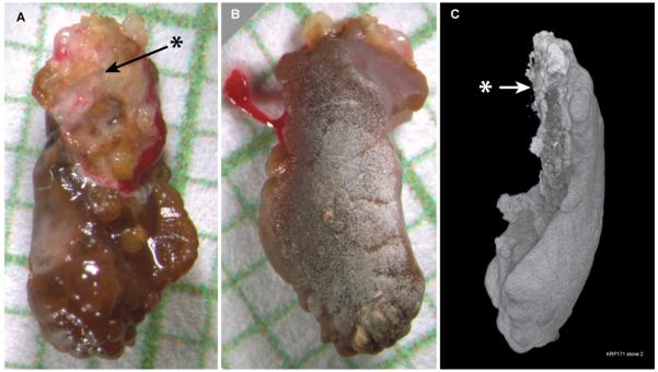
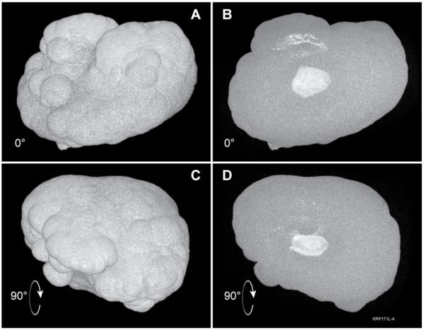
Patients and methods: ICSF undergoing percutaneous nephrolithotomy or ureteroscopy for the treatment of nephrolithiasis gave consent to participate in this study. All accessible renal papillae were endoscopically imaged using a digital endoscope. All stones were removed and determined by the operating surgeon to be attached or unattached to the underlying papilla. Micro-computed tomography (micro-CT), which provides three-dimensional analysis of entire stones, was used to compare the structure and composition of attached and unattached stones.
Results: Of 115 stones collected from nine patients (12 renal units), only 25 stones were found not to be attached to renal papillae. Of these 25 stones, four were lost and 12 showed definite morphological evidence of having been attached to tissue, probably having been displaced from papillae during access. For the remaining nine stones, micro-CT analysis showed at least one internal region of calcium phosphate within each of these unattached CaOx stones, i.e. the internal structure of the unattached stones is consistent with their having originated attached to Randall's plaque, and then having become detached but retained in the kidney, with new layers of CaOx eventually covering the original attachment site. CONCLUSIONS; Micro-CT analysis supports the hypothesis that in ICSF, both attached and unattached stones occur as a result of a common pathogenic mechanism, i.e. in this type of stone former, CaOx stones, even those not showing morphology that betrays attachment, all originate attached to interstitial plaque on the renal papilla.
Figures

References
-
- Randall A. The etiology of primary renal calculus. International Abstract of Surgery. 1940;71:209–40.
-
- Evan AP, Lingeman J, Coe FL, Worcester E. Randall’s plaque: Pathogenesis and role in calcium oxalate nephrolithiasis. Kidney Int. 2006;69:1313–8. - PubMed
-
- Evan AP, Coe FL, Lingeman JE, Shao Y, Sommer AJ, Bledsoe SB, et al. Mechanism of formation of human calcium oxalate renal stones on Randall’s plaque. Anat Rec. 2007;290:1315–23. - PubMed
Publication types
MeSH terms
Substances
Grants and funding
LinkOut - more resources
Full Text Sources
