. 2009 Aug;110(4):1226-40.
doi: 10.1111/j.1471-4159.2009.06212.x. Epub 2009 Jun 15.

The HDAC inhibitor, sodium butyrate, stimulates neurogenesis in the ischemic brain

Affiliations

The HDAC inhibitor, sodium butyrate, stimulates neurogenesis in the ischemic brain

Hyeon Ju Kim et al. J Neurochem. 2009 Aug.

Abstract

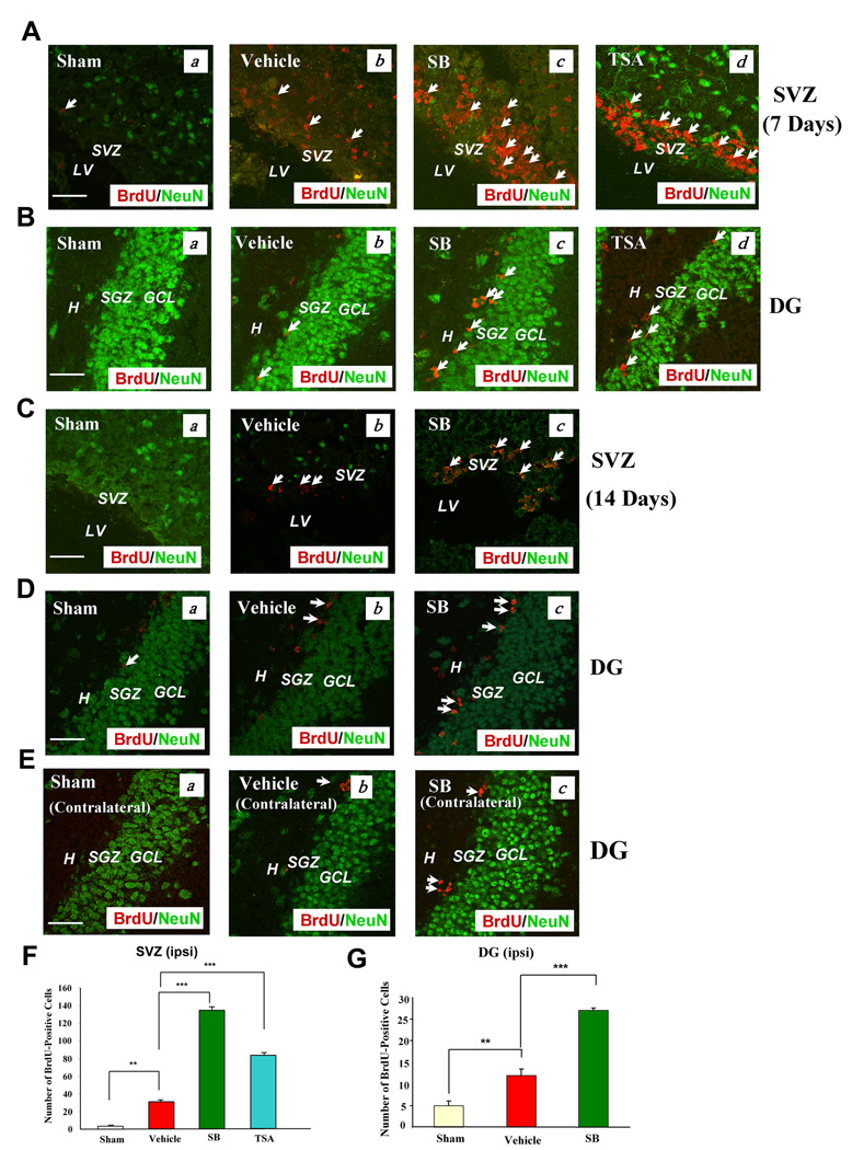
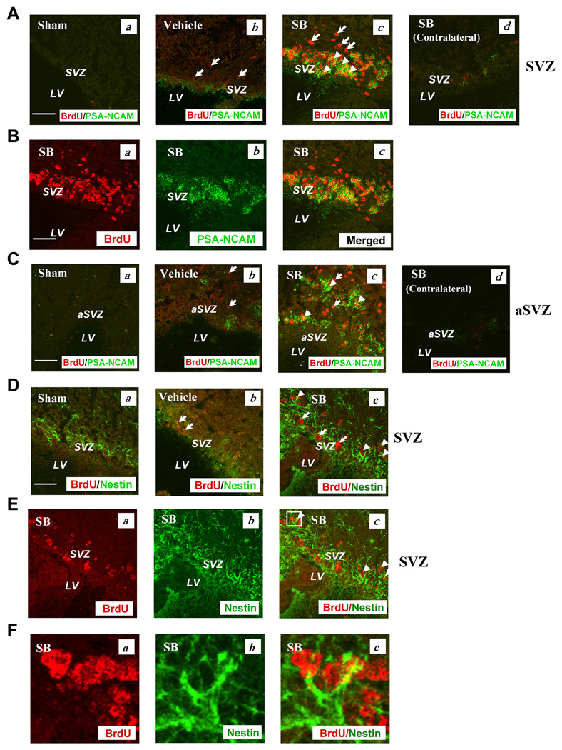
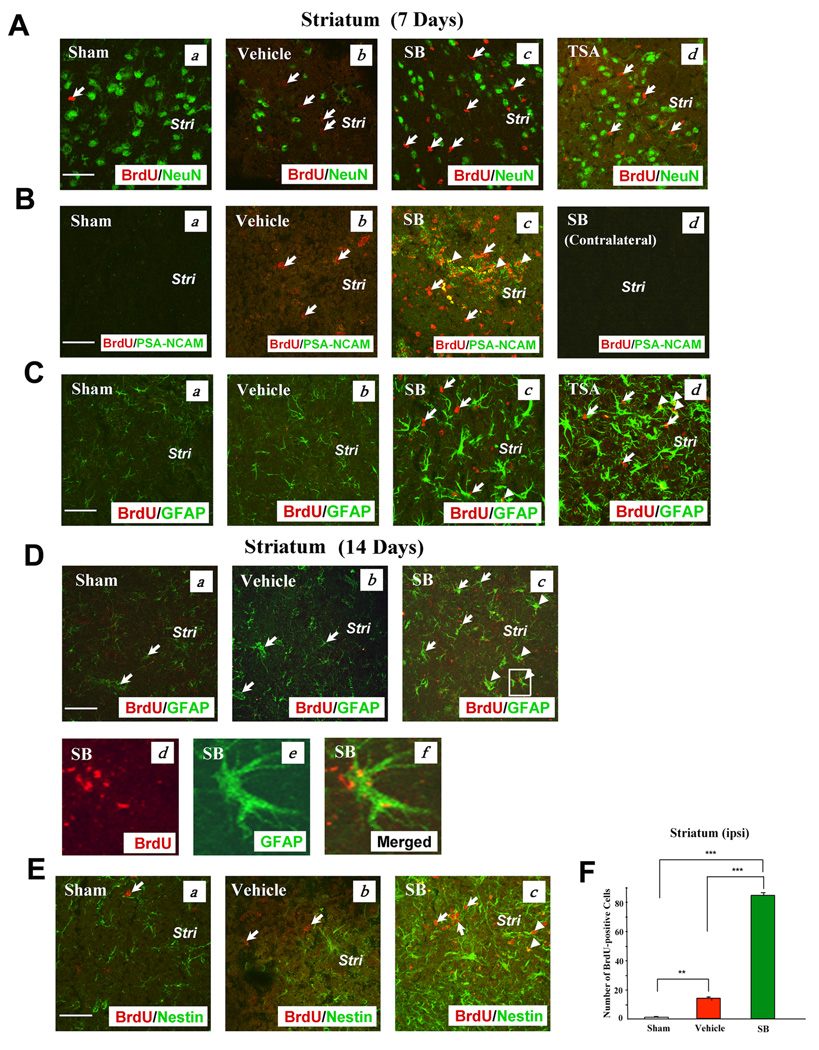
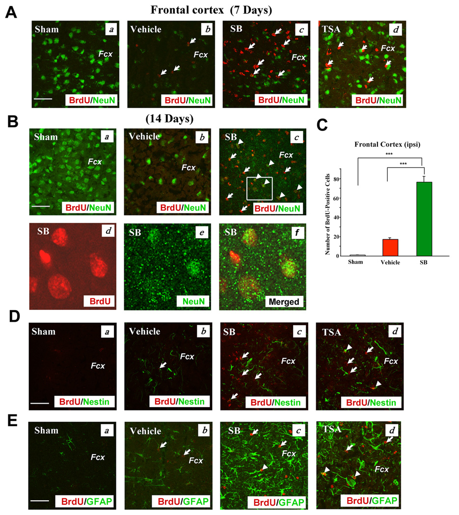
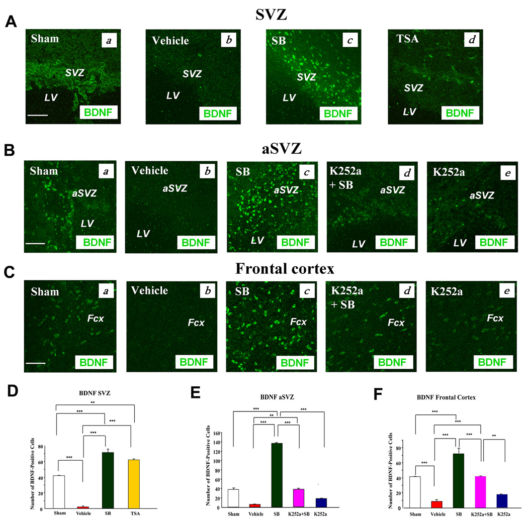
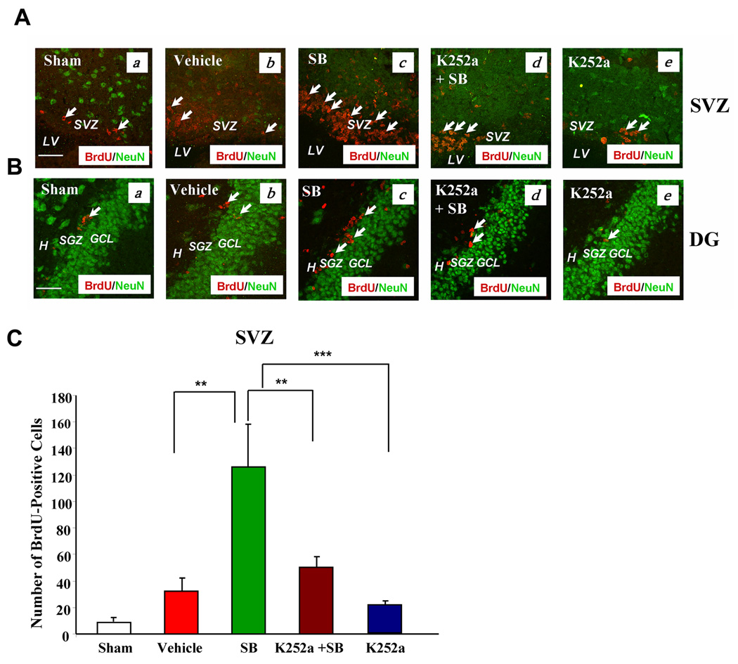
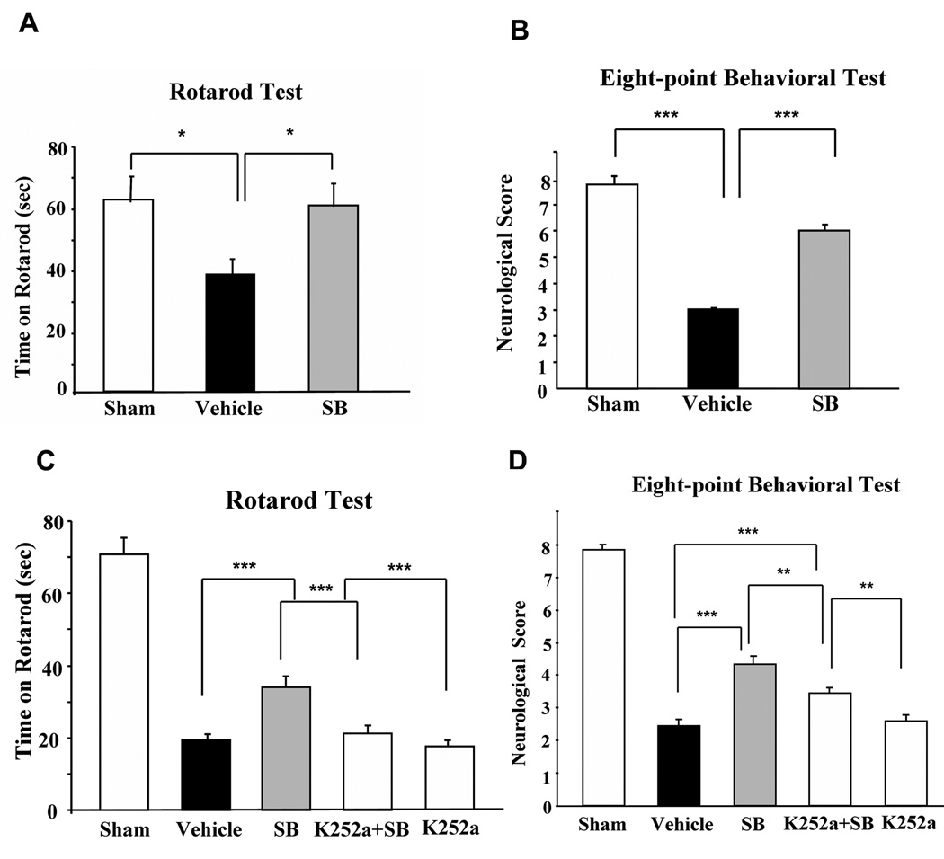
In the healthy adult brain, neurogenesis normally occurs in the subventricular zone (SVZ) and hippocampal dentate gyrus (DG). Cerebral ischemia enhances neurogenesis in neurogenic and non-neurogenic regions of the ischemic brain of adult rodents. This study demonstrated that post-insult treatment with a histone deacetylase inhibitor, sodium butyrate (SB), stimulated the incorporation of bromo-2'-deoxyuridine (BrdU) in the SVZ, DG, striatum, and frontal cortex in the ischemic brain of rats subjected to permanent cerebral ischemia. SB treatment also increased the number of cells expressing polysialic acid-neural cell adhesion molecule, nestin, glial fibrillary acidic protein, phospho-cAMP response element-binding protein (CREB), and brain-derived neurotrophic factor (BDNF) in various brain regions after cerebral ischemia. Furthermore, extensive co-localization of BrdU and polysialic acid-neural cell adhesion molecule was observed in multiple regions after ischemia, and SB treatment up-regulated protein levels of BDNF, phospho-CREB, and glial fibrillary acidic protein. Intraventricular injection of K252a, a tyrosine kinase B receptor antagonist, markedly reduced SB-induced cell proliferation detected by BrdU and Ki67 in the ipsilateral SVZ, DG, and other brain regions, blocked SB-induced nestin expression and CREB activation, and attenuated the long-lasting behavioral benefits of SB. Together, these results suggest that histone deacetylase inhibitor-induced cell proliferation, migration and differentiation require BDNF-tyrosine kinase B signaling and may contribute to long-term beneficial effects of SB after ischemic injury.


Figures

Fig. 1
Fig. 1. Post pMCAO-insult treatment of rats with SB increased the number of BrdU (+) cells in the SVZ and DG of the ischemic hemisphere
Brain tissues were analyzed by double immunostaining with BrdU (red) and NeuN (green, a mature neuronal marker). (A & B) Seven days post pMCAO treatment. (a) Sham-operated, (b) vehicle-treated, pMCAO, (c) SB-treated, pMCAO, (d) TSA-treated, pMCAO. Arrows identify BrdU (+) cells. Scale bar = 50 µm. The vehicles for SB and TSA were saline and DMSO respectively; no significant differences were found in stimulating cell proliferation. (C, D & E) Representative data analyzed from 3–4 animals per group, 14 days post pMCAO treatment. (a) Sham, (b) vehicle, (c) SB. Arrows identify BrdU (+) cells. Scale bar = 50 µm. Fewer BrdU (+) cells were found in the contralateral hemisphere than in the ipsilateral hemisphere (E). (F & G) Quantified results of the number of BrdU (+) cells in the ipsilateral SVZ on day 7, and DG on Day 14 post-pMCAO, respectively. Data are mean ± SEM (n = 3–4 animals per group). **p<0.01, ***p<0.001, between indicated groups. SVZ: subventricular zone, LV: lateral ventricle. DG: dentate gyrus, H: hilus, SGZ: subgranular zone, GCL: granular cell layer.
Fig. 2
Fig. 2. SB treatment markedly increased the number of cells expressing PSA-NCAM in the SVZ on Day 7 post pMCAO
Brain tissues were analyzed by double immunostaining with BrdU (red) and PSA-NCAM (green). (A) (a) Sham, (b) vehicle, (c) SB, (d) SB in the contralateral SVZ. (B) Most BrdU (+) cells showed colocalization with PSA-NCAM in the SVZ (yellow). (a) BrdU (+) cells in the SB-treated group, (b) PSA-NCAM (+) cells in the SB-treated group, (c) merged image. (C) SB treatment increased PSA-NCAM immunostaining in the ipsilateral aSVZ. (a) Sham, (b) vehicle, (c) SB, (d) SB in the contralateral aSVZ. (D) Nestin, a neuroblast marker, was increased by SB treatment in the ipsilateral SVZ on Day 14 post pMCAO. (a) Sham, (b) vehicle, (c) SB. (E) (a) BrdU (+) cells in the SB-treated group, (b) nestin (+) cells in the SB group, (c) merged image. (F) Enlarged images from the box in Fig. 2E (c). Arrows identify BrdU (+) cells. Arrowheads identify cells with colocalized expression of BrdU/PSA-NCAM or BrdU/nestin. Scale bar = 50 µm. SVZ: subventricular zone, aSVZ: anterior subventricular zone, LV: lateral ventricle.
Fig. 3
Fig. 3. SB or TSA treatment robustly increased BrdU, PSA-NCAM, GFAP, and nestin-positive immunostaining in the ipsilateral striatum on Days 7 or 14 after pMCAO
(A) BrdU (+) cells and NeuN (+) cells did not show colocalization on Day 7 after pMCAO. (a) Sham, (b) vehicle, (c) SB, (d) TSA in the corresponding areas of the striatum. BrdU (red), NeuN (green). Arrows identify BrdU (+) cells. (B) BrdU and PSA-NCAM were both extensively present on Day 7 after pMCAO. (a) Sham, (b) vehicle, (c) SB in the corresponding areas, (d) SB in the contralateral striatum. BrdU (red), PSA-NCAM (green), colocalized BrdU and PSA-NCAM (yellow). Arrows identify BrdU (+) cells. Arrowheads identify cells with colocalized expression of BrdU and PSA-NCAM. (C) GFAP (+) cells were markedly increased by SB or TSA on Day 7 after pMCAO. (a) Sham, (b) vehicle, (c) SB, (d) TSA. BrdU (red), GFAP (green), colocalized BrdU and GFAP (yellow). Arrows identify BrdU (+) cells. Arrowheads identify cells with colocalized expression of BrdU and GFAP. (D) SB treatment increased GFAP (+) cells on Day 14 following cerebral ischemia. (a) Sham, (b) vehicle, (c–f) SB. BrdU (red), GFAP (green), colocalized BrdU and GFAP (yellow). Arrows identify GFAP (+) cells. Arrowheads identify cells with colocalized expression of BrdU and GFAP. (d–f) enlarged area indicated in (c). (E) Nestin was potentiated by SB treatment on Day 14 after pMCAO. (a) Sham, (b) vehicle, (c) SB. BrdU (red), nestin (green), colocalized BrdU and nestin (yellow). Arrows identify BrdU (+) cells. Arrowheads identify cells with colocalized expression of BrdU and nestin. Stri: striatum. Scale bar = 50 µm. (F) Quantification of BrdU (+) cells in the ipsilateral striatum of Sham-operated, Vehicle-treated and SB-treated rats on day 7 following pMCAO. Data are mean ± SEM (n = 3–4 animals per group). **p<0.01, ***p<0.001, between indicated groups.
Fig. 4
Fig. 4. SB treatment robustly increased BrdU, NeuN, PSA-NCAM, GFAP, and nestin-positive immunostaining in the frontal cortex of the ischemic hemisphere on Day 7 or 14 after pMCAO
(A) Double immunostaining of BrdU (+) and NeuN (+) cells on Day 7 after stroke. (a) Sham, (b) vehicle, (c) SB, (d) TSA in the corresponding areas of the frontal cortex. BrdU (red), NeuN (green). Arrows identify BrdU (+) cells. (B) Day 14 after cerebral ischemia. BrdU was coexpressed with NeuN in some cells. (a) Sham, (b) vehicle, (c) SB, (d–f) enlarged area indicated in (c). BrdU (red), NeuN (green), colocalized BrdU and NeuN (yellow). Arrows identify BrdU (+) cells. Arrowheads identify cells with colocalized expression of BrdU and NeuN. (C) Quantification of BrdU (+) cells in the ipsilateral frontal cortex of Sham-operated, Vehicle-treated and SB-treated rats on day 7 following pMCAO. Data are mean ± SEM (n = 3–4 animals per group). ***p<0.001, between indicated groups. (D) Immunostaining of BrdU (+) and nestin (+) cells on Day 7 following cerebral ischemia. (a) Sham, (b) vehicle, (c) SB, (d) TSA. BrdU (red), nestin (green). Arrows identify BrdU (+) cells. Arrowheads identify cells with colocalized expression of BrdU and nestin. Fcx: frontal cortex, Scale bar = 50 µm. Data obtained from representative confocal microscopy sections of rat brains (n = 3–4, each group). (E) Double immunostaining of BrdU (+) and GFAP (+) cells on Day 14 after ischemia. (a) Sham, (b) vehicle, (c) SB, (d) TSA. BrdU (red), GFAP (green), colocalized BrdU and GFAP (yellow). Arrows identify BrdU (+) cells. Arrowheads identify cells with colocalized expression of BrdU and GFAP.
Fig. 5
Fig. 5. BDNF immunoreactivity was markedly down-regulated in the vehicle-treated group, but up-regulated in SB- or TSA-treated rats in the ipsilateral SVZ, aSVZ, and frontal cortex on Day 7 after pMCAO
SB-induced BDNF up-regulation in the aSVZ and frontal cortex was blocked by co-treatment with K252a. (A) (a) Sham, (b) vehicle, (c) SB, (d) TSA. BDNF (green). Scale bar = 50 µm. Results are from representative brain sections (n = 3–4 per group). (B) (a) Sham, (b) vehicle, (c) SB, (d) K252a + SB, (e) K252a. BDNF (green). Scale bar = 50 µm. (C) (a) Sham, (b) vehicle, (c) SB, (d) K252a + SB, (e) K252a. BDNF (green). Scale bar = 50 µm. Experimental conditions are described in the Methods. SVZ: subventricular zone, LV: lateral ventricle, aSVZ: anterior subventricular zone, Fcx: frontal cortex. (D), (E) & (F) Quantified results of BDNF (+) cells in the SVZ, aSVZ and frontal cortex under the experimental conditions shown in (A)–(C). Data are mean ± SEM and were analyzed from 4 rats per group, **p< 0.01, ***p< 0.001, between indicated groups.
Fig. 6
Fig. 6. Intracerebral injection of K252a into the lateral ventricle robustly suppressed BrdU labels in the SVZ and DG of the ipsilateral hemispheres on Day 7 following cerebral ischemia
(A) (a) Sham, (b) vehicle, (c) SB, (d) K252a + SB, (e) K252a groups in the SVZ. BrdU (red), NeuN (green). Arrows identify BrdU (+) cells. (B) (a) Sham, (b) vehicle, (c) SB, (d) K252a + SB, (e) K252a groups in the DG. BrdU (red), NeuN (green). Arrows identify BrdU (+) cells. Scale bar = 50 µm. (C) Quantification of the number of BrdU-labeled cells from the ipsilateral SVZ in sham operated, vehicle, SB, K252a + SB, and K252a-treated rats on Day 7 following pMCAO. Data are mean ± SEM., **p< 0.01, ***p<0.001, comparison between the indicated groups. Data were analyzed from 3–4 animals per group. SVZ: subventricular zone, LV: lateral ventricle. H: hilus, SGZ: subgranular zone, GCL: granular cell layer.
Fig. 7
Fig. 7. Long-term behavioral benefits of SB in pMCAO rats were sensitive to K252a treatment
(A & B) Post-insult SB treatment largely prevented ischemia-induced neurological deficits as determined on Day 14 after pMCAO by rotarod (A) and an eight-point behavioral test (B). Data were analyzed from sham-operated animals (n = 5), vehicle (normal saline)-treated (n = 6), and SB-treated animals (n = 6). Data are mean ± SEM. *p< 0.05, and ***p<0.001 compared with the vehicle group. (C & D) K252a attenuated the beneficial effects of SB as determined by rotarod (C) and eight-point behavioral test (D) on Day 7 after pMCAO. Data were obtained from sham-operated animals (n = 7), vehicle (normal saline)-treated (n = 9), SB-treated (n = 9), K252a + SB-treated (n = 9), and K252a-treated animals (n = 10). Data are mean ± SEM. **p< 0.01, and ***p<0.001, comparison between the indicated groups.

References

    1. Alvarez-Buylla A, Garcia-Verdugo JM. Neurogenesis in adult subventricular zone. J. Neurosci. 2002;22:629–634. - PMC - PubMed
    1. Arvidsson A, Collin T, Kirik D, Kokaia Z, Lindvall O. Neural replacement from endogenous precursors in the adult brain after stroke. Nat. Med. 2002;8:963–970. - PubMed
    1. Barnabé-Heider F, Miller FD. Endogenously produced neurotrophins regulate survival and differentiation of cortical progenitors via distinct signaling pathways. J. Neurosci. 2003;23:5149–5160. - PMC - PubMed
    1. Benraiss A, Chmielnicki E, Lerner K, Roh D, Goldman SA. Adenoviral brain derived neurotrophic factor induces both neostriatal and olfactory neuronal recruitment from endogenous progenitor cells in the adult forebrain. J. Neurosci. 2001;21:6718–6731. - PMC - PubMed
    1. Bonfanti L. PSA-NCAM in mammalian structural plasticity and neurogenesis. Prog. Neurobiol. 2006;80:129–164. - PubMed

Publication types

MeSH terms