. 2009 Jun 23:6:84.
doi: 10.1186/1743-422X-6-84.

Hepatitis C virus core, NS3, NS4B and NS5A are the major immunogenic proteins in humoral immunity in chronic HCV infection

Affiliations

Hepatitis C virus core, NS3, NS4B and NS5A are the major immunogenic proteins in humoral immunity in chronic HCV infection

Maarit Sillanpää et al. Virol J. .

Abstract

Background: The viral genome of hepatitis C virus constitutes a 9.6-kb single-stranded positive-sense RNA which encodes altogether 11 viral proteins. In order to study the humoral immune responses against different HCV proteins in patients suffering from chronic HCV infection, we produced three structural (core, E1 and E2) and six nonstructural proteins (NS2, NS3, NS4A, NS4B, NS5A and NS5B) in Sf9 insect cells by using the baculovirus expression system.

Results: The recombinant HCV core, E1, E2, NS2, NS3, NS4A, NS4B, NS5A and NS5B proteins were purified and used in Western blot analysis to determine antibody responses against individual HCV protein in 68 HCV RNA and antibody positive human sera that were obtained from patients suffering from genotype 1, 2, 3 or 4 infection. These sera were also analysed with INNO-LIA Score test for HCV antibodies against core, NS3, NS4AB and NS5A, and the results were similar to the ones obtained by Western blot method. Based on our Western blot analyses we found that the major immunogenic HCV antigens were the core, NS4B, NS3 and NS5A proteins which were recognized in 97%, 86%, 68% and 53% of patient sera, respectively. There were no major genotype specific differences in antibody responses to individual HCV proteins. A common feature within the studied sera was that all except two sera recognized the core protein in high titers, whereas none of the sera recognized NS2 protein and only three sera (from genotype 3) recognised NS5B.

Conclusion: The data shows significant variation in the specificity in humoral immunity in chronic HCV patients.


Figures

Figure 1
Figure 1
Expression of recombinant HCV proteins. A. Individual HCV genes were cloned into pcDNA3.1(+) plasmid under CMV promoter. The expression of HCV proteins was verified by in vitro translation. The proteins were metabolically labeled with [35S]-methionine and separated on 15% (core, E1, NS4A) or on 12% (E2, NS2, NS3, NS5A, NS5B, NS4B) SDS-PAGE and autoradiographed. B. SDS-PAGE analysis of purified recombinant HCV proteins. Individual HCV genes were inserted into baculovirus expression plasmids and recombinant HCV-expressing baculoviruses were obtained. Recombinant HCV proteins were produced in Sf9 cells followed by purification of the proteins by preparative SDS-PAGE. Samples of purified HCV proteins (0.5–1 μg each) were separated on 15% (core, E1 and NS4A) or 12% (E2, NS3, NS5A, NS5B, NS2 and NS4B) SDS-PAGE and stained by Coomassie Blue.
Figure 2
Figure 2
Detection of anti-HCV antibodies in HCV RNA and antibody positive human sera with recombinant HCV proteins. 1 μg of each baculovirus-expressed and preparative SDS-PAGE-purified recombinant HCV protein was loaded onto 10–20% Tris-glycine polyacrylamide gradient gels. Core, NS2, NS3, NS4A, NS4B, and NS5A were loaded on one gel, and E1, E2, and NS5B on another gel, respectively. 3 μg of Sf9 cell extract was also loaded onto one gel as a control. Proteins separated on gels were transferred to nylon membranes, sliced and stained with serially diluted human serum obtained from HCV RNA and antibody positive patients. The following dilutions were used (lane 1) 1:100, (lane 2) 1:500, (lane 3) 1:2500, (lane 4) 1:12500 and 1:62500 (not shown). After incubation with secondary Abs, the bands were visualized by 3-amino-9-ethylcarbazole (AEC). A. An example of highly positive human serum number 36 is shown, B. an example of a weakly positive human serum number 17 is shown.
Figure 3
Figure 3
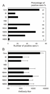
The specificity of anti-HCV antibody responses in patients suffering from chronic HCV infection. A. The frequency of antibodies against individual recombinant HCV proteins in 68 serum specimens obtained from patients suffering from chronic HCV infection. Both the number and percentage of positive sera are shown. B. The relative antibody levels against individual HCV proteins were determined as the last serum dilution showing a positive signal in Western blot analysis. The means and standard deviations of the means for antibody levels against individual HCV proteins are shown based on 68 HCV RNA and antibody positive patient sera. Only individuals showing a positive antibody response against a given HCV protein are included into the means.
Figure 4
Figure 4
Anti-HCV antibody responses in patients infected with different HCV genotypes. The mean antibody titers and standard deviations against individual recombinant HCV proteins when samples were grouped according to the HCV genotypes 1, 2, 3 or 4. The analysis contained 21 sera of genotype 1, 20 sera of genotype 2, 23 sera of genotype 3 and 4 sera of genotype 4. Only those serum specimens showing a positive response to a given HCV protein are included in the means.
Figure 5
Figure 5
The persistence of anti-HCV antibody responses against individual HCV proteins. HCV antibody prevalence in five HCV RNA and antibody positive individuals infected with genotype 1b or 3a was analyzed from serum specimens obtained before and after IFN-α monotherapy (black bar). Relative anti-HCV antibody titers were determined by Western blot analysis as described in Fig. 2.

References

    1. Walewski JL, Keller TR, Stump DD, Branch AD. Evidence for a new hepatitis C virus antigen encoded in an overlapping reading frame. RNA. 2001;7:710–721. doi: 10.1017/S1355838201010111. - DOI - PMC - PubMed
    1. Lindenbach BD, Rice CM. Unravelling hepatitis C virus replication from genome to function. Nature. 2005;436:933–938. doi: 10.1038/nature04077. - DOI - PubMed
    1. Xu Z, Choi J, Yen TS, Lu W, Strohecker A, Govindarajan S, Chien D, Selby MJ, Ou J. Synthesis of a novel hepatitis C virus protein by ribosomal frameshift. EMBO J. 2001;20:3840–3848. doi: 10.1093/emboj/20.14.3840. - DOI - PMC - PubMed
    1. Griffin SD, Beales LP, Clarke DS, Worsfold O, Evans SD, Jaeger J, Harris MP, Rowlands DJ. The p7 protein of hepatitis C virus forms an ion channel that is blocked by the antiviral drug, Amantadine. FEBS Lett. 2003;535:34–38. doi: 10.1016/S0014-5793(02)03851-6. - DOI - PubMed
    1. Jones CT, Murray CL, Eastman DK, Tassello J, Rice CM. Hepatitis C virus p7 and NS2 proteins are essential for production of infectious virus. J Virol. 2007;81:8374–8383. doi: 10.1128/JVI.00690-07. - DOI - PMC - PubMed

Publication types

MeSH terms

Substances

LinkOut - more resources