Full-length cytokeratin-19 is released by human tumor cells: a potential role in metastatic progression of breast cancer
- PMID: 19549321
- PMCID: PMC2716508
- DOI: 10.1186/bcr2326
Full-length cytokeratin-19 is released by human tumor cells: a potential role in metastatic progression of breast cancer
Abstract
Introduction: We evaluated whether CK19, one of the main cytoskeleton proteins of epithelial cells, is released as full-length protein from viable tumor cells and whether this property is relevant for metastatic progression in breast cancer patients.
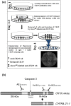
Methods: EPISPOT (EPithelial ImmunoSPOT) assays were performed to analyze the release of full-length CK19 by carcinoma cells of various origins, and the sequence of CK19 was analyzed with mass spectrometry. Additional functional experiments with cycloheximide, Brefeldin A, or vincristine were done to analyze the biology of the CK19-release. CK19-EPISPOT was used to detect disseminated tumor cells in bone marrow (BM) of 45 breast cancer patients who were then followed up over a median of 6 years.
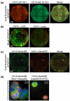
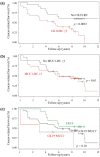
Results: CK19 was expressed and released by colorectal (HT-29, HCT116, Caco-2) and breast (MCF-7, SKBR3, and MDA-MB-231) cancer cell lines. The CK19-EPISPOT was more sensitive than the CK19-ELISA. Dual fluorescent EPISPOT with antibodies against different CK19 epitopes showed the release of the full-length CK19, which was confirmed by mass spectrometry. Functional experiments indicated that CK19 release was an active process and not simply the consequence of cell death. CK19-releasing cells (RCs) were detectable in BM of 44% to 70% of breast cancer patients. This incidence and the number of CK19-RCs were correlated to the presence of overt metastases, and patients with CK19-RCs had a reduced survival as compared with patients without these cells (P = 0.025, log-rank test; P = 0.0019, hazard ratio, 4.7; multivariate analysis).
Conclusions: Full-length CK19 is released by viable epithelial tumor cells, and CK19-RCs might constitute a biologically active subset of breast cancer cells with high metastatic properties.
Figures


References
-
- Fuchs E, Weber K. Intermediate filaments: structure, dynamics, function, and disease. Annu Rev Biochem. 1994;63:345–382. - PubMed
-
- Coulombe PA, Omary MB. 'Hard' and 'soft' principles defining the structure, function and regulation of keratin intermediate filaments. Curr Opin Cell Biol. 2002;14:110–122. - PubMed
-
- Dohmoto K, Hojo S, Fujita J, Yang Y, Ueda Y, Bandoh S, Yamaji Y, Ohtsuki Y, Dobashi N, Ishida T, Takahara J. The role of caspase 3 in producing cytokeratin 19 fragment (CYFRA21-1) in human lung cancer cell lines. Int J Cancer. 2001;91:468–473. - PubMed
-
- Chu PG, Weiss LM. Keratin expression in human tissues and neoplasms. Histopathology. 2002;40:403–439. - PubMed
-
- Pujol JL, Grenier J, Daures JP, Daver A, Pujol H, Michel FB. Serum fragment of cytokeratin subunit 19 measured by CYFRA 21-1 immunoradiometric assay as a marker of lung cancer. Cancer Res. 1993;53:61–66. - PubMed
Publication types
MeSH terms
Substances
LinkOut - more resources
Full Text Sources
Other Literature Sources
Medical
Miscellaneous

