. 2009 Jul 15;122(Pt 14):2554-66.
doi: 10.1242/jcs.046250. Epub 2009 Jun 23.

Caspase cleavage of Atg4D stimulates GABARAP-L1 processing and triggers mitochondrial targeting and apoptosis

Affiliations

Caspase cleavage of Atg4D stimulates GABARAP-L1 processing and triggers mitochondrial targeting and apoptosis

Virginie M S Betin et al. J Cell Sci. .

Abstract

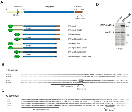
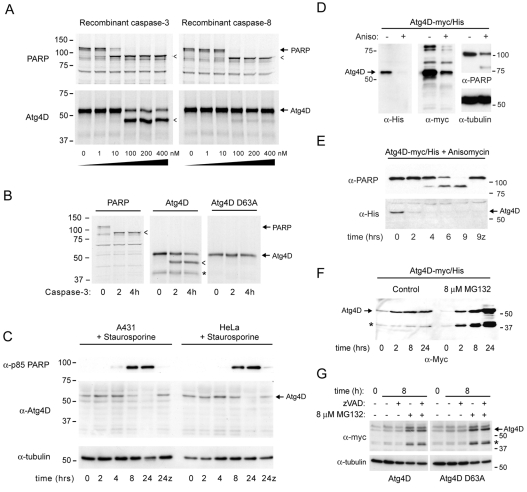
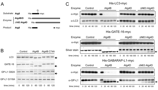
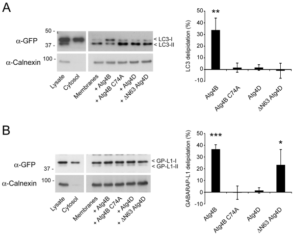
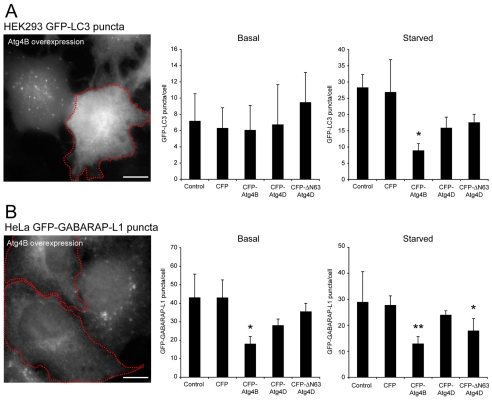
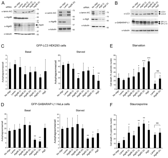
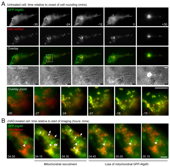
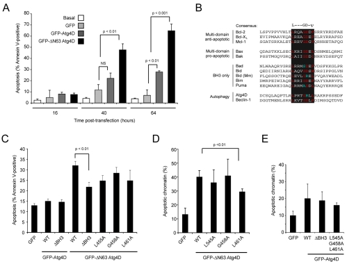
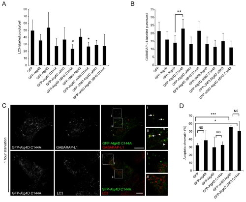
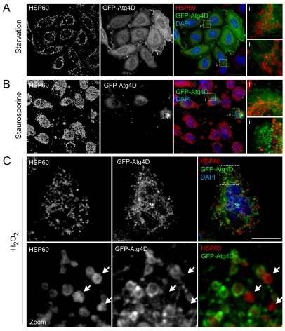
Autophagy is an important catabolic process with roles in cell survival and cell death. It sequesters cytosol and organelles within double-membrane autophagosomes that deliver their contents to lysosomes for degradation. Autophagosome biogenesis is coordinated by the autophagy-related protein 4 (Atg4) family of C54 endopeptidases (Atg4A-Atg4D). These enzymes prime and then later delipidate the autophagosome marker, Atg8. Here, we show that one family member, Atg4D, is cleaved by caspase-3 in vitro and in apoptotic cells. Atg4D is a poor priming and delipidation enzyme in vitro, but truncated DeltaN63 Atg4D displays increased activity against the Atg8 paralogue, gamma-aminobutyric acid receptor-associated protein-like 1 (GABARAP-L1). In living cells, DeltaN63 Atg4D stimulates the delipidation of GABARAP-L1, whereas siRNA silencing of the gene expressing Atg4D abrogates GABARAP-L1 autophagosome formation and sensitises cells to starvation and staurosporine-induced cell death. Interestingly, Atg4D overexpression induces apoptosis, which is preceded by the caspase-independent recruitment of Atg4D to mitochondria and is facilitated by a putative C-terminal Bcl-2 homology 3 (BH3) domain. Atg4D also acquires affinity for damaged mitochondria in cells treated with hydrogen peroxide. These data suggest that Atg4D is an autophagy regulator that links mitochondrial dysfunction with apoptosis.


Figures

Fig. 1.
Fig. 1.
Domain structure and alignment of human autophagins, and expression of Atg4D. (A) Domain structure of human Atg4D showing the location of the features described in parts (B) and (C), and cartoons depicting the various Atg4D constructs used in this study. C144 is an essential cysteine in the active site triad of Atg4D. (B) Alignment of the N-termini of the four presumed, human Atg4 paralogues (Atg4A-Atg4D). Atg4C and Atg4D have extended N-termini, containing putative caspase cleavage motifs (DEVD). (C) Alignment of the C-termini of human Atg4A-Atg4D. Atg4D contains a putative BH3 domain towards the end of the molecule (see text for details). (D) Immunoblot of HeLa cell extracts using polyclonal anti-Atg4D antiserum. Control and GFP-Atg4D transfected cell lysates are shown. A band at the expected molecular mass of the endogenous protein (∼54 kDa) is indicated, as is a ∼78 kDa band representing GFP-Atg4D.
Fig. 2.
Fig. 2.
Caspase cleavage and cellular turnover of Atg4D. (A) 35S-labelled human Atg4D translated in vitro and subjected to caspase cleavage using increasing concentration of recombinant caspase-3 and caspase-8 (90 minutes at 30°C). Autoradiographs of SDS-PAGE gels are shown. Human PARP is shown as a positive control. Cleavage products are indicated by arrowheads. (B) Site-directed mutagenesis of the aspartate at position 63 (D63A) prevented caspase cleavage of Atg4D. 35S-labelled, recombinant wild type and caspase-resistant (D63A) Atg4D incubated for increasing times with 100 nM caspase-3. Rapid cleavage of wild type but not D63A was seen (arrowhead). An additional band was observed at all time-points in the wild-type Atg4D samples (*). (C) Cleavage of Atg4D in living cells undergoing staurosporine-induced apoptosis. Human A431cells (to the left) and HeLa cells (to the right) were incubated with 1 μM staurosporine for increasing lengths of time, and extracts were immunoblotted for caspase-cleaved p85 PARP and Atg4D. Tubulin is shown as a loading control. By 24 hours, no Atg4D was detected, and this was prevented by the caspase inhibitor zVAD.fmk (z). (D-G) Proteasome-mediated turnover of human Atg4D. (D) HeLa cells transiently expressing myc/His-tagged Atg4D (Atg4D-myc/His) were incubated with anisomycin (6 hours), and extracts were immunoblotted with antibodies recognising myc, His, PARP and α-tubulin. The signal for Atg4D was diminished in anisomycin-treated samples. (E) HeLa cells transiently expressing Atg4D-myc/His were treated with anisomycin for up to 9 hours in the absence or presence of zVAD.fmk (z), and immunoblotted for PARP and Atg4D. Loss of Atg4D-myc/His preceded PARP cleavage and was not prevented by zVAD.fmk. (F-G) HeLa cells transiently expressing wild-type or caspase-resistant (G) Atg4D-myc/His were incubated in the absence or presence of the proteasome inhibitor MG132. MG132 caused the accumulation of full-length and ∼42 kDa Atg4D (*) both in wild-type cells and in cells expressing caspase-resistant Atg4D, in the absence or presence of zVAD.fmk.
Fig. 3.
Fig. 3.
Action of Atg4D and ΔN63 Atg4D in Atg8 priming assays. (A) Schematic of the reagents used for in vitro assays. Atg8 paralogues were double-tagged (6xHis-Atg8-myc). (B) Priming efficiency of Atg4B and its C74A active-site mutant against LC3, GATE-16 and GABARAP-L1 (G92A and G116A mutants). Arrows mark the positions of primed products. (C) In vitro Atg8 priming assays. Recombinant Atg8 paralogues were incubated with Atg4B, Atg4D and ΔN63 Atg4D and analysed by immunoblotting and/or by silver staining. Priming products are indicated by arrows.
Fig. 4.
Fig. 4.
Action of Atg4D and ΔN63 Atg4D in Atg8 delipidation assays. Autophagosome-enriched membranes from cells stably expressing YFP-LC3 (A) and GFP-GABARAP-L1 (B) were incubated with recombinant Atg4B (wild type and C74A), Atg4D and ΔN63 Atg4D, and analysed by immunoblotting with anti-GFP antibodies. Lipidated LC3 (LC3-II) and GABARAP-L1 (GP-L1-II) migrated faster than the delipidated products (LC3-I and GP-L1-I, respectively). Graphs show means ± s.d. of three independent experiments. Student's t-test: *P<0.05, **P<0.01, *** P<0.001.
Fig. 5.
Fig. 5.
Overexpression supports a role for ΔN63 Atg4D in GABARAP-L1 processing in living cells. (A) HEK293 cells stably expressing GFP-LC3 and (B) HeLa cells stably expressing GFP-GABARAP-L1 were transiently transfected with CFP, CFP-Atg4B, CFP-Atg4D or CFP-ΔN63 Atg4D. Bar charts show autophagosome numbers (mean ± s.d. of three independent experiments; n>50 cells for each treatment) assessed under basal and starvation conditions (see Materials and Methods). Example images of GFP-LC3 and GFP-GABARAP-L1 puncta are shown to the left for Atg4B-expressing cells (red outline; scale bar: 10 μm). Additional representative images are shown in supplementary material Fig. S3. Student's t-test for values compared with CFP controls: *P<0.05, **P<0.01.
Fig. 6.
Fig. 6.
siRNA-mediated Atg4D silencing sensitises cells to starvation and cell death. (A) Immunoblots of HeLa cells silenced for Atg4D, Atg4B and Atg5. Silencing was achieved over 72 hours and lamin was included as a negative control. An additional oligonucleotide for Atg4B is included (centre panel) because of inconsistencies in the responses of cells to Atg4B knockdown (see text). (B) Immunoblots of HeLa cells treated with the oligonucleotides indicated for 72 hours and subjected to starvation (1 hour). Lipidated Atg8 paralogues (LC3-II; GP-L1-II) migrate slightly further in SDS-PAGE. (C,D) Atg4 silencing abrogates GABARAP-L1 autophagosomes formation. HEK293 cells stably expressing GFP-LC3 (C) and HeLa cells stably expressing GFP-GABARAP-L1 (D) were depleted for Lamin, Atg4B, Atg4D or Atg5 (72 hours), then subjected or not to starvation (1 hour) before being fixed and analysed for autophagosome numbers. Silencing Atg4D but not Atg4B restricted GABARAP-L1 puncta formation. Silencing Atg5, Atg4B (one oligonucleotide from three) and Atg4D (one oligonucleotide from two) significantly reduced LC3 autophagosome formation in starved cells. Bar charts shows means ± s.d. of three independent experiments (150-450 cells for each treatment). (E) Depletion of Atg5 and Atg4D sensitises cells to starvation-induced cell death. HeLa cells were silenced as indicated (72 hours), then incubated for 5 hours in starvation media. (F) Depletion of Atg5 and Atg4D sensitised cells to staurosporine-induced cell death. HeLa cells were silenced as indicated (72 hours), then incubated for 5 hours with 1 μM staurosporine. For parts (E) and (F), cell death was assessed by DAPI fluorescence, scoring for pyknotic chromatin. Means ± s.d. are shown for three independent experiments. Student's t-test for values compared to the lamin controls: *P=0.5, **P=0.01, ***P=0.001.
Fig. 7.
Fig. 7.
GFP-Atg4D is recruited to mitochondria as a prelude to apoptotic cell death. (A) Time-lapse analysis of an A431 cell transiently co-expressing GFP-Atg4D (green) and Mito-dsRed (red). GFP-Atg4D was rapidly recruited to mitochondria ∼24 minutes before cell rounding and apoptosis (note, yellow signal in the zoomed panels). These sequences were taken from supplementary material Movie 1. Scale bars: 10 μm. (B) Caspase activity is not required for mitochondrial recruitment of GFP-Atg4D. Time-lapse analysis of mitochondrial GFP-Atg4D recruitment in a zVAD.fmk-treated A431 cell. Note the transient appearance of rings of GFP-Atg4D (green) in proximity to mitochondria (red) (arrows). Centrosomal GFP-Atg4D is indicated by an arrowhead. These sequences were taken from supplementary material Movie 2. Scale bar: 10 μm.
Fig. 8.
Fig. 8.
Assessment of Atg4D-induced cytotoxicity. (A) Overexpression of Atg4D and ΔN63 Atg4D triggers apoptosis. HeLa cells expressing GFP, GFP-Atg4D or GFP-ΔN63 Atg4D were labelled with fluorescent annexin V at 16, 40 and 64 hours post-transfection. Means ± s.d. are shown for three independent experiments with the P-values for Student's t-test. (B-E) Atg4D contains a putative C-terminal BH3 domain that facilitates overexpression-induced apoptosis. (B) Alignment of BH3 domains from Bcl-2 family members with pro- or anti-apoptotic roles, Beclin-1 and Atg4D (Mm, Mus musculus). (C) Analysis of apoptosis in cells overexpressing GFP-Atg4D, GFP-ΔN63 Atg4D and BH3 mutants by annexin-V-binding (40 hours). (D,E) Analysis of apoptosis in cells overexpressing GFP-Atg4D, GFP-ΔN63 Atg4D and BH3 mutants by scoring pyknotic chromatin (40-hour expression). Data show means ± s.d. of three independent experiments, scored blind. P-values for Student's t-test are shown.
Fig. 9.
Fig. 9.
Dissociation of Atg4D-mediated autophagy from overexpression-induced apoptosis. (A,B) HeLa cells transiently expressing the constructs shown (refer to Fig. 1A) were fixed and labelled with anti-LC3 antibodies (A) or GABARAP-L1 antibodies (B). Autophagosome numbers were counted in three independent experiments (>100 cells; bars represent means ± s.d.) In (A), means are compared with GFP control; *P<0.5 using the Student's t-test. (C) Inactive Atg4D colocalises with a population of GABARAP-L1-positive puncta. Confocal images are shown of starved (1 hour) HeLa cells transiently expressing GFP-Atg4D C144A, fixed and stained with antibodies against LC3 and GABARAP-L1. GFP-positive puncta were observed and these colocalised with a population of GABARAP-L1-positive puncta (arrows in zoom panels) but not with LC3 puncta (GABARAP-L1 puncta not co-labelled with GFP-Atg4D C144A are indicated by arrowheads). Scale bar: 10 μm. (D) Atg4D endopeptidase activity is not required for overexpression-induced apoptosis. HeLa cells transiently overexpressing (64 hours) GFP-Atg4B, GFP-Atg4D, GFP-ΔN63 Atg4D and Atg4D C144A constructs (see Fig. 1A) were scored for pyknotic chromatin. Data show means ± s.d. of three independent experiments. Student's t-test: *P<0.5, **P<0.01, ***P<0.001. NS, not significant.
Fig. 10.
Fig. 10.
Analysis of Atg4D localisation in response to cellular stress. HeLa cells transiently expressing GFP-Atg4D were starved (A), treated with staurosporine (1 μM) (B) or treated with hydrogen peroxide (10 nM) (C) for 5 hours, then fixed and labelled with antibodies against HSP60. In a sub-population of starved cells, small GFP-Atg4D puncta were observed in the vicinity of mitochondria (Aii); however, no evidence of mitochondrial Atg4D localisation was observed in starved or staurosporine-treated cells (A,B). Note that in late apoptotic cells Atg4D formed aggregates, but these did not colocalise with mitochondria (Bii; see also supplementary material Movie 1). (C) GFP-Atg4D relocates to mitochondria in a sub-population of H2O2-treated HeLa cells. Mitochondria not labelled with GFP-Atg4D are indicated with arrows. In parts (A) and (B) zoomed panels are shown to the right. Scale bars: 10 μm (A,B), 5 μm (C).

References

    1. Axe, E. L., Walker, S. A., Manifava, M., Chandra, P., Roderick, H. L., Habermann, A., Griffiths, G. and Ktistakis, N. T. (2008). Autophagosome formation from membrane compartments enriched in phosphatidylinositol 3-phosphate and dynamically connected to the endoplasmic reticulum. J. Cell Biol. 182, 685-701. - PMC - PubMed
    1. Baehrecke, E. H. (2005). Autophagy: dual roles in life and death? Nat. Rev. Mol. Cell Biol. 6, 505-510. - PubMed
    1. Botti, J., Djavaheri-Mergny, M., Pilatte, Y. and Codogno, P. (2006). Autophagy signaling and the cogwheels of cancer. Autophagy 2, 67-73. - PubMed
    1. Broustas, C. G., Gokhale, P. C., Rahman, A., Dritschilo, A., Ahmad, I. and Kasid, U. (2004). BRCC2, a novel BH3-like domain-containing protein, induces apoptosis in a caspase-dependent manner. J. Biol. Chem. 279, 26780-26788. - PubMed
    1. Chen, G., Cizeau, J., Vande Velde, C., Park, J. H., Bozek, G., Bolton, J., Shi, L., Dubik, D. and Greenberg, A. (1999). Nix and Nip3 form a subfamily of pro-apoptotic mitochondrial proteins. J. Biol. Chem. 274, 7-10. - PubMed

Publication types

MeSH terms