. 2009 Jul 7;106(27):11090-5.
doi: 10.1073/pnas.0903535106. Epub 2009 Jun 22.

Structure and assembly of immature HIV

Affiliations

Structure and assembly of immature HIV

J A G Briggs et al. Proc Natl Acad Sci U S A. .

Abstract

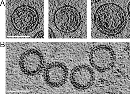
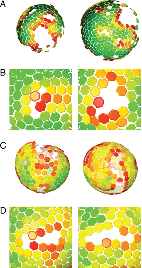
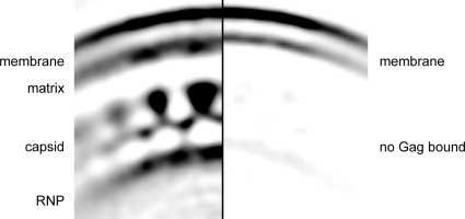
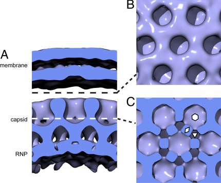
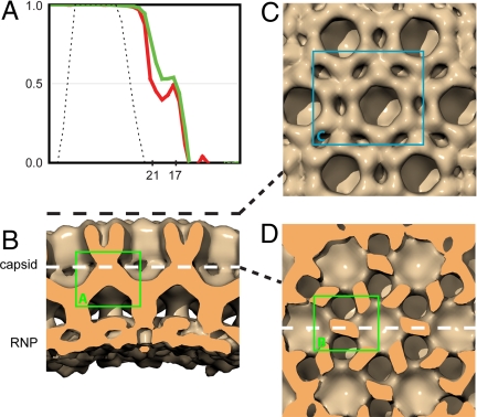
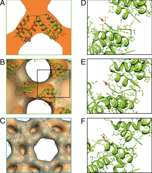
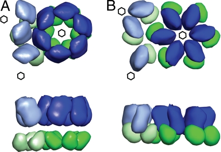
The major structural components of HIV are synthesized as a 55-kDa polyprotein, Gag. Particle formation is driven by the self-assembly of Gag into a curved hexameric lattice, the structure of which is poorly understood. We used cryoelectron tomography and contrast-transfer-function corrected subtomogram averaging to study the structure of the assembled immature Gag lattice to approximately 17-A resolution. Gag is arranged in the immature virus as a single, continuous, but incomplete hexameric lattice whose curvature is mediated without a requirement for pentameric defects. The resolution of the structure allows positioning of individual protein domains. High-resolution crystal structures were fitted into the reconstruction to locate protein-protein interfaces involved in Gag assembly, and to identify the structural transformations associated with virus maturation. The results of this study suggest a concept for the formation of nonsymmetrical enveloped viruses of variable sizes.


Conflict of interest statement

The authors declare no conflict of interest.

Figures

Fig. 1.
Fig. 1.
Sections through tomograms of (A) immature virus particles and (B) in-vitro-assembled Gag particles. Scale bar, 100 nm.
Fig. 2.
Fig. 2.
Global lattice maps of HIV particles. Positions of hexameric unit cells are marked with hexamers. Hexamers are colored according to cross-correlation on a scale from low (red) to high (green). Maps are shown in perspective such that hexamers on the rear surface of the particle appear smaller. (A) Lattice maps for immature HIV particles. The side of the particle toward the viewer lacks ordered Gag. (B) Close-up of defects in immature Gag lattice. (C) Lattice maps for in-vitro-assembled Gag particles. (D) Close-up of defects in in-vitro-assembled particle lattice.
Fig. 3.
Fig. 3.
Part of central section through reconstruction of immature virus particle from regions with Gag bound to the membrane (Left) or where no Gag is bound to the membrane (Right). Density is dark on a light background. The inner leaflet of the bilayer is thicker in regions where Gag is present, but the bilayer spacing remains constant.
Fig. 4.
Fig. 4.
Surface rendering of reconstruction of immature virus particle. (A) Surface cut perpendicular to the membrane to reveal the 2 membrane leaflets, the CA layer, and the RNP. (B) Surface cut tangential to the membrane at a radius indicated by the dashed black line in A, and looking down on the N-CA domains. (C) Surface cut tangentially along the white dashed line in A, through the C-CA domains. Hexagon, triangle, and rhombus indicate examples of 6-fold, 3-fold, and 2-fold symmetry axes, respectively.
Fig. 5.
Fig. 5.
Reconstruction of in-vitro-assembled Gag particles. (A) Fourier shell correlation plot, before (red) and after (green) CTF correction. The black dotted line indicates the band-pass filter applied during the reconstruction process. The resolutions measured according to the 0.5 criterion are 21 Å before CTF correction, and 17 Å after CTF correction. (B) Surface rendering sectioned perpendicular to the particle surface. (C) Surface cut tangential to the surface at a radius indicated by the dashed black line in B, looking down on the N-CA domains. (D) Surface cut tangentially along the white dashed line in B, through the C-CA domains. Colored boxes refer to the regions illustrated in Fig. 6 A–C.
Fig. 6.
Fig. 6.
Fitting crystal structures into the in vitro particle reconstruction. (A) The best identified fit of the C-CA dimer PDB3ds2 into the region boxed in Fig. 5B. (B) The same fit viewed as boxed in Fig. 5D. (C) One possible fit of the N-CA domain, viewed as boxed in Fig. 5C. (D and E) Close-ups of the inter-dimer contacts highlighted with the black box in 6B. (D–F) The fits shown are for Dimer 2, PDB1a8o (D), Dimer 3, PDB3ds2 (E), and Dimer 4, PDB2ont (F). Red arrows indicate the position of the N-terminal residue of C-CA.
Fig. 7.
Fig. 7.
Schematic of the structural changes taking place during maturation. (A) The immature arrangement of the N-terminal (blue) and C-terminal (green) domains of CA, viewed from outside the particle (Top), and rotated 90 ° around the horizontal axis (Bottom). Domains from neighboring hexamers are indicated in lighter colors. Six-fold lattice positions are marked by hexagons. (B) The mature arrangement, with domains positioned according to ref. .

References

    1. Demirov DG, Freed EO. Retrovirus budding. Virus Res. 2004;106:87–102. - PubMed
    1. Morita E, Sundquist WI. Retrovirus budding. Annu Rev Cell Dev Biol. 2004;20:395–425. - PubMed
    1. Wilk T, et al. Organization of immature human immunodeficiency virus type 1. J Virol. 2001;75:759–771. - PMC - PubMed
    1. Briggs JAG, Johnson MC, Simon MN, Fuller SD, Vogt VM. Cryo-electron microscopy reveals conserved and divergent features of Gag packing in immature particles of Rous Sarcoma virus and human immunodeficiency virus. J Mol Biol. 2006;355:157–168. - PubMed
    1. Adamson CS, Freed EO. Human immunodeficiency virus type 1 assembly, release, and maturation. Adv Pharmacol. 2007;55:347–387. - PubMed

Publication types

Substances

LinkOut - more resources