. 2009 Jul 1;69(13):5357-63.
doi: 10.1158/0008-5472.CAN-08-3895. Epub 2009 Jun 23.

Nanoscale cellular changes in field carcinogenesis detected by partial wave spectroscopy

Affiliations

Nanoscale cellular changes in field carcinogenesis detected by partial wave spectroscopy

Hariharan Subramanian et al. Cancer Res. .

Abstract

Understanding alteration of cell morphology in disease has been hampered by the diffraction-limited resolution of optical microscopy (>200 nm). We recently developed an optical microscopy technique, partial wave spectroscopy (PWS), which is capable of quantifying statistical properties of cell structure at the nanoscale. Here we use PWS to show for the first time the increase in the disorder strength of the nanoscale architecture not only in tumor cells but also in the microscopically normal-appearing cells outside of the tumor. Although genetic and epigenetic alterations have been previously observed in the field of carcinogenesis, these cells were considered morphologically normal. Our data show organ-wide alteration in cell nanoarchitecture. This seems to be a general event in carcinogenesis, which is supported by our data in three types of cancer: colon, pancreatic, and lung. These results have important implications in that PWS can be used as a new method to identify patients harboring malignant or premalignant tumors by interrogating easily accessible tissue sites distant from the location of the lesion.


Figures
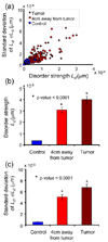
Figure 1
Figure 1
(a) Cells at a distance from a colon tumor undergo changes in their internal nanoarchitecture similar to tumor cells. The values of Ld and its intracellular standard deviation σLd averaged over a cell, that is Ld(c) and σ(c), for control cells, tumor cells, and cells at a 4 cm distance from tumor plotted in Ld(c), σ(c) parameter space. Each point in this diagram corresponds to a single cell. As can be seen, the histologically normal cells at a distance from a tumor have an increased disorder strength due to field carcinogenesis. (b) The relative values of the disorder strength Ld(d) for the control cells, tumor cells and cells away from tumor. Ld(g) is obtained by averaging Ld(c) from randomly chosen ~30 cells for each of the three cell types. The error bar represents the standard error of the mean. The average disorder strength Ld(g) is significantly increased in the tumor cells compare to control cells (Student t-test p-value <0.0001). More importantly, cells 4 cm away from tumor also undergo increased disorder strength compared to control (p-value < 0.0001). (c) The values of the intracellular standard deviation of the disorder strength σ(g) for the three cell types. σ(g) is obtained from ~30 cells for each cell types. The error bars represent the standard errors of the means. σ(g) is progressively increased in cells 4 cm away from tumor to the tumor cells compared to control (all p-values < 0.0001).
Figure 2
Figure 2
Cells obtained from histologically normal colonic mucosa have increased disorder strength due to the presence of premalignant tumors anywhere else in colon. (a) The values Ld(c) and σ(c) obtained from histologically normal rectal mucosa from control patients and those with adenomatous polyp elsewhere in the colon. Though histologically normal, the rectal cells from patients with premalignant tumors occupy a separate regime in the parameter space with only slight overlap with the normal cells. (b) The relative values of the disorder strength Ld(g) for cells from histologically normal rectal mucosa in patients with premalignant tumors and those with no tumors present. The average disorder strength Ld(g) is significantly elevated in cells from patients with presence of adenomatous polyps elsewhere in the colon compared to controls (p-value <0.0001). (c) The relative values of intracellular standard deviation of the disorder strength σ(g) from histologically normal rectal mucosa. σ(g) is obtained from ~30 cells for each cell types. The error bars represent the standard errors of the means. Similar to Ld(g), the σ(g) is significantly elevated in patients with the presence of premalignant tumors elsewhere in colon (p-values < 0.0001).
Figure 3
Figure 3
(a) Histologically normal duodenal mucosa cells have increased disorder strength due to the presence of pancreatic cancer. The Ld(c) and σ(c) obtained from histologically normal duodenal mucosa from patients with pancreatic cancer and with no cancer. Though histologically normal, the Ld(c) and σ(c) from the duodenal cells are significantly elevated in patients with pancreatic cancer (p-value < 0.0001) and the values only slightly overlap with the cells from control patients. (b) Cells obtained from histologically normal buccal mucosa have increased disorder strength due to the presence of lung cancer. The values of Ld(c) and σ(c) obtained from histologically normal buccal mucosa from patients with chronic obstructive pulmonary disease COPD and those with lung cancer. Though histologically normal, the buccal mucosa cells have a much higher Ld(c) and σ(c) in patients with lung cancer compared to those patients with COPD (p-value < 0.0001).

Similar articles

Cited by

References

    1. Lewis JD, Ng K, Hung KE, et al. Detection of proximal adenomatous polyps with screening sigmoidoscopy: a systematic review and meta-analysis of screening colonoscopy. Arch Intern Med. 2003;163:413–420. - PubMed
    1. Takayama T, Katsuki S, Takahashi Y, et al. Aberrant crypt foci of the colon as precursors of adenoma and cancer. N Engl J Med. 1998;339:1277–1284. - PubMed
    1. Anti M, Marra G, Armelao F, et al. Rectal epithelial cell proliferation patterns as predictors of adenomatous colorectal polyp recurrence. Gut. 1993;34:525–530. - PMC - PubMed
    1. Bernstein C, Bernstein H, Garewal H, et al. A bile acid-induced apoptosis assay for colon cancer risk and associated quality control studies. Cancer Res. 1999;59:2353–2357. - PubMed
    1. Chen L, Hao C, Chiu Y, et al. Alteration of Gene Expression in Normal-Appearing Colon Mucosa of APCmin Mice and Human Cancer Patients. Cancer Research. 2004;64:3694–3700. - PubMed

Publication types