. 2009 Jul 1;69(13):5312-20.
doi: 10.1158/0008-5472.CAN-08-3767. Epub 2009 Jun 23.

Active Notch1 confers a transformed phenotype to primary human melanocytes

Affiliations

Active Notch1 confers a transformed phenotype to primary human melanocytes

Chelsea C Pinnix et al. Cancer Res. .

Abstract

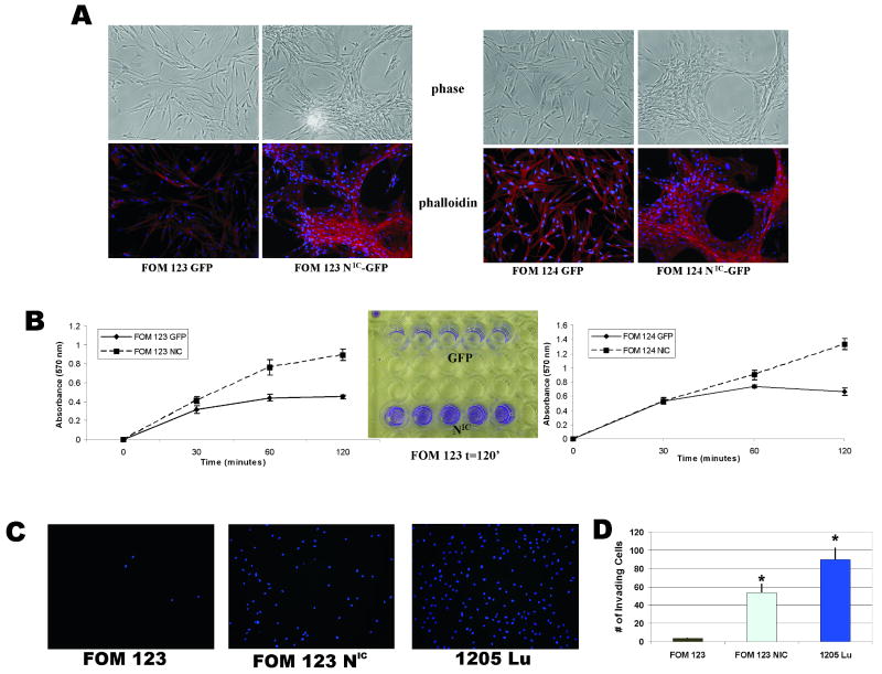
The importance of mitogen-activated protein kinase signaling in melanoma is underscored by the prevalence of activating mutations in N-Ras and B-Raf, yet clinical development of inhibitors of this pathway has been largely ineffective, suggesting that alternative oncogenes may also promote melanoma. Notch is an interesting candidate that has only been correlated with melanoma development and progression; a thorough assessment of tumor-initiating effects of activated Notch on human melanocytes would clarify the mounting correlative evidence and perhaps identify a novel target for an otherwise untreatable disease. Analysis of a substantial panel of cell lines and patient lesions showed that Notch activity is significantly higher in melanomas than their nontransformed counterparts. The use of a constitutively active, truncated Notch transgene construct (N(IC)) was exploited to determine if Notch activation is a "driving" event in melanocytic transformation or instead a "passenger" event associated with melanoma progression. N(IC)-infected melanocytes displayed increased proliferative capacity and biological features more reminiscent of melanoma, such as dysregulated cell adhesion and migration. Gene expression analyses supported these observations and aided in the identification of MCAM, an adhesion molecule associated with acquisition of the malignant phenotype, as a direct target of Notch transactivation. N(IC)-positive melanocytes grew at clonal density, proliferated in limiting media conditions, and also exhibited anchorage-independent growth, suggesting that Notch alone is a transforming oncogene in human melanocytes, a phenomenon not previously described for any melanoma oncogene. This new information yields valuable insight into the basic epidemiology of melanoma and launches a realm of possibilities for drug intervention in this deadly disease.


Figures

Figure 1
Figure 1. Notch signaling pathway activation status in melanocytes and melanoma cells
A, Immunohistochemical analysis of active Notch1 expression in primary human melanoma lesions. Anti-HMB45 was employed to identify cells of melanocytic origin. Darker staining depicts melanin production whereas active Notch1 and HMB45 are indicated by red staining. Unaffected epidermal and dermal tissue immediately adjacent to the primary melanoma lesion was also photographed for control purposes. B, Quantitative RT-PCR analysis of Notch1 expression in a panel of melanoma lesions and cell lines. Fold change is reflected against a representative human melanocyte cell line, HEMN. C, (Top) Microarray analysis of Hes1, Hey1 and Hey2 expression in four laser microdissected melanoma lesions, four melanoma cell lines, and four primary melanocyte cell lines. (Bottom) Microarray analysis of Hes1, Hey1 and Hey2 expression in a panel of human melanoma cell lines. D, Western blot analysis of activated Notch1 protein in melanocytes and melanoma cells from C.
Figure 2
Figure 2. Inhibition of Notch signaling suppresses melanoma, but not melanocyte growth in vitro
A, Expression of DN-MAML protein in melanoma cell line WM 3248 was detected by immunoblotting for the myc-tagged DN-MAML. Effect of DN-MAML expression on Notch1 targets Hes1, Hey1 and Hey2 was determined by quantitative RT-PCR in 2 melanoma cell lines, WM3248 and WM1366. B, 7 day growth curves of melanocytes and melanoma cells infected with DN-MAML or GFP lentiviruses. * p<0.05 (Student's T-test). C, Growth inhibition in melanocytes and melanoma cells in the presence of increasing concentrations of a γ-secretase inhibitor. Cell growth was determined by MTT analysis. Results are percentage of growth inhibition compared with untreated controls (adjusted to 0%). * p<0.005 (Student's t-test).
Figure 3
Figure 3. Constitutive activation of the Notch1 pathway enhances melanocyte growth in vitro
A, (Left) mRNA analysis of Notch1 expression in GFP- and NIC-infected FOM 117 cells by RT-PCR. (Right) Western blot analysis of Notch1 protein expression in GFP and NIC-infected FOM 117 cells. B, Immunofluorescence analysis of NIC localization in FOM 117 GFP and NIC-infected cells. C, mRNA expression of Hey1 and Hey2 in FOM 117 GFP and NIC-infected cells as determined by quantitative RT-PCR. D, ∼ 2 week growth curve of GFP- and NIC-infected FOM 123 and 124 cells. *p<0.005 (Student's T-test).
Figure 4
Figure 4. Constitutive Notch1 activation induces cytoskeletal, adhesion, and migratory changes in primary melanocytes
A, Phalloidin immunostaining reveals morphological changes induced by active Notch1 expression. FOM 123 and 124 NIC cells form networks of capillary like structures when grown in monolayer as compared to the normal dendritic growth pattern of normal GFP-infected cells. B, Cell adhesion assay demonstrated that NIC-infected FOM cells possess increased adhesive properties compared to GFP-infected controls within 2 hours post-plating. *p<0.005 (Student's t-test). C & D, NIC- and GFP-infected melanocytes were subject to Boyden chamber assays to test their ability to migrate through matrigel. After 24 hours, membranes stained with DAPI demonstrated enhanced ability of NIC-infected FOM cells to migrate; 1205Lu melanoma cells acted as a positive control.
Figure 5
Figure 5. Identification of a CSL binding sequence within the human MCAM promoter
A, Immunoblot analysis of E-cadherin, MCAM, β3-integrin, and active Notch1 protein levels in primary melanocytes infected with GFP (G) or NIC (N) lentiviruses. B, FOM 117 melanocytes were plated on Jagged1-Fc or control Fc plates for 72 hours and subsequently assayed for active Notch1 and MCAM expression by immunoblotting. C, Enhanced MCAM protein expression in NIC-infected WM 3248 melanoma cells (left). Interruption of Notch signaling by treatment with 5 μM GSI X decreased MCAM expression in uninfected WM 3248 cells, as determined by immunoblotting for MCAM (right). D, (Top) Multiple consensus binding sequences for CSL, the hexamer TGGGAA, were identified within the human MCAM promoter at positions -477 and -3500 (see text for details). (Bottom) Chromatin immunoprecipitation (ChIP) analysis demonstrating association of CSL with regions of the MCAM promoter. Hes1 acts as a positive control for binding activity, while MCAM -1.5K is a negative control.
Figure 6
Figure 6. Notch1 activation transforms primary melanocytes in vitro
A, Constitutive Notch1 activation induces focus formation in NIC-infected melanocytes grown in monolayer on tissue culture dishes. *p<0.005 (Student's T-test). B, NIC-infected melanocytes display increased survival in limiting growth factor conditions, as depicted. 100% media indicates normal melanocytes media containing bFGF, SCF, and ET3. 10% media is a 10-fold decrease in FBS; the growth factor(s) present in each respective medium is described beneath each panel. *p<0.005 (Student's T-test). C, NIC-infected melanocytes display anchorage-independent growth in soft agar. *p<0.005 (Student's T-test).

References

    1. Artavanis-Tsakonas S, Rand MD, Lake RJ. Notch signaling: cell fate control and signal integration in development. Science (New York, NY. 1999;284:770–6. - PubMed
    1. Ellisen LW, Bird J, West DC, et al. TAN-1, the human homolog of the Drosophila notch gene, is broken by chromosomal translocations in T lymphoblastic neoplasms. Cell. 1991;66:649–61. - PubMed
    1. Shou J, Ross S, Koeppen H, de Sauvage FJ, Gao WQ. Dynamics of notch expression during murine prostate development and tumorigenesis. Cancer Res. 2001;61:7291–7. - PubMed
    1. Raafat A, Bargo S, Anver MR, Callahan R. Mammary development and tumorigenesis in mice expressing a truncated human Notch4/Int3 intracellular domain (h-Int3sh) Oncogene. 2004;23:9401–7. - PubMed
    1. Sriuranpong V, Borges MW, Ravi RK, et al. Notch signaling induces cell cycle arrest in small cell lung cancer cells. Cancer Res. 2001;61:3200–5. - PubMed

Publication types

MeSH terms