. 2009 Jul 15;69(14):5743-51.
doi: 10.1158/0008-5472.CAN-08-3564. Epub 2009 Jun 23.

Breast cancer migration and invasion depend on proteasome degradation of regulator of G-protein signaling 4

Affiliations

Breast cancer migration and invasion depend on proteasome degradation of regulator of G-protein signaling 4

Yan Xie et al. Cancer Res. .

Abstract

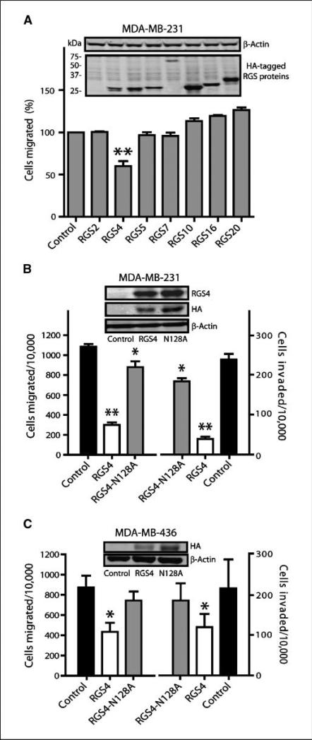
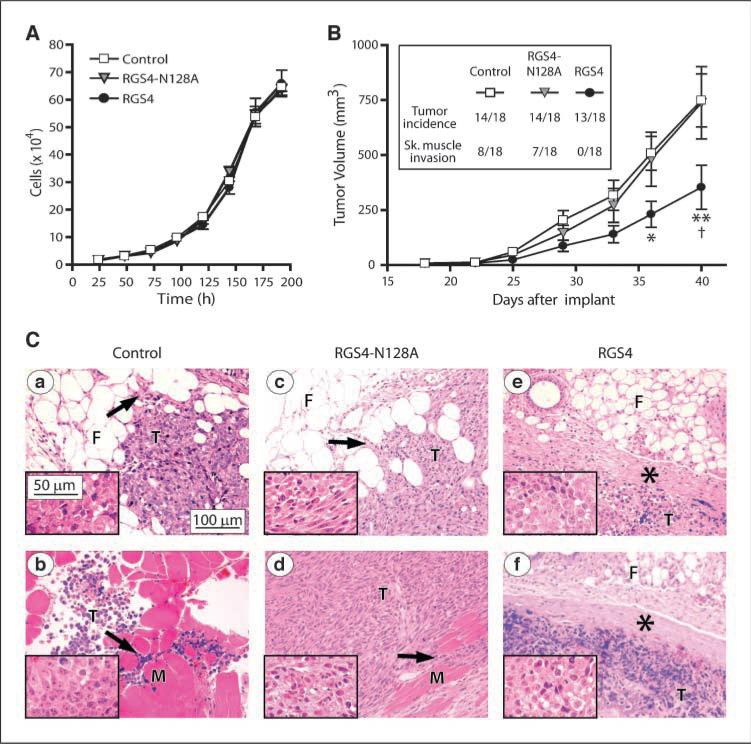
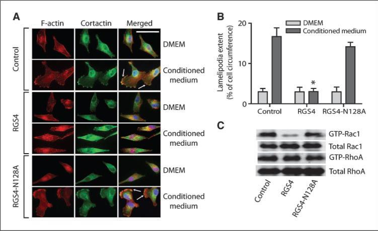
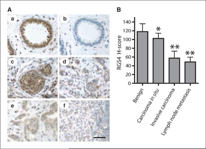
Aberrant signaling through G-protein coupled receptors promotes metastasis, the major cause of breast cancer death. We identified regulator of G-protein signaling 4 (RGS4) as a novel suppressor of breast cancer migration and invasion, important steps of metastatic cascades. By blocking signals initiated through G(i)-coupled receptors, such as protease-activated receptor 1 and CXC chemokine receptor 4, RGS4 disrupted Rac1-dependent lamellipodia formation, a key step involved in cancer migration and invasion. RGS4 has GTPase-activating protein (GAP) activity, which inhibits G-protein coupled receptor signaling by deactivating G-proteins. An RGS4 GAP-deficient mutant failed to inhibit migration and invasion of breast cancer cells in both in vitro assays and a mouse xenograft model. Interestingly, both established breast cancer cell lines and human breast cancer specimens showed that the highest levels of RGS4 protein were expressed in normal breast epithelia and that RGS4 down-regulation by proteasome degradation is an index of breast cancer invasiveness. Proteasome blockade increased endogenous RGS4 protein to levels that markedly inhibit breast cancer cell migration and invasion, which was reversed by an RGS4-targeted short hairpin RNA. Our findings point to the existence of a mechanism for posttranslational regulation of RGS4 function, which may have important implications for the acquisition of a metastatic phenotype by breast cancer cells. Preventing degradation of RGS4 protein should attenuate aberrant signal inputs from multiple G(i)-coupled receptors, thereby retarding the spread of breast cancer cells and making them targets for surgery, radiation, and immune treatment.


Figures

Figure 1
Figure 1
Exogenously expressed RGS4 protein inhibits breast cancer cell migration and invasion. Columns, mean; bars, SE with *, P < 0.05 and **, P < 0.01 compared with control cells. A, comparison of Transwell migration of MDA-MB-231 cells transfected with HA-tagged RGS proteins normalized to vector controls (n = 6, 100% = 755 ± 98 per 10,000 seeded cells). Transwell migration and invasion of MDA-MB-231 cells stably expressing RGS4 or RGS4-N128A (B, n = 5) or MDA-MB-436 cells transiently expressing RGS4 or RGS4-N128A (C, n = 3). Insets, Western blot of HA-tagged RGS proteins and β-actin.
Figure 2
Figure 2
RGS4 inhibits breast tumor growth and suppresses tumor invasion in nude mice. A, in vitro growth of MDA-MB-231 control cells or cells stably expressing RGS4 or RGS4-N128A. Points, mean (n = 4 triplicate experiments); bars, SE. B, in vivo tumor growth derived from MDA-MB-231 cell variants injected into mouse mammary fat pads (n = 18 mice per group; inset, tumor incidence and skeletal muscle invasion). Points, mean for formed tumors; bars, SE. *, P < 0.05 and **, P < 0.001 compared with control, †, P < 0.001 compared with RGS4-N128A using two-way ANOVA with Bonferroni comparison of means. C, representative H&E-stained micrographs of tumor tissue from mice injected with MDA-MB-231 control cells showing (a) mammary fat pad and (b) skeletal muscle invasion, RGS4-N128A–expressing cells showing (c) mammary fat pad and (d) skeletal muscle invasion, and RGS4-expressing cells showing well demarcated tumor boundaries (e and f, asterisks). Arrows, infiltration of the tumor cells (T) into the fibroadipose tissue (F) or skeletal muscle (M). Insets, higher magnification images of the tumor cells.
Figure 3
Figure 3
RGS4 selectively inhibits Rac-activated lamellipodia formation in breast cancer cells. A and B, RGS4 blocks lamellipodia formation and the translocation of cortactin to lamellipodia by inhibiting Rac activity in MDA-MB-231 cells. Arrows, lamellipodia at cell edges (A). Scale bar, 50 μm. B, lamellipodial extent at cell leading edges was quantified as fraction of cell circumference on 20 randomly selected cells in each group. Columns, means; bars, SE (n = 3), *, P < 0.001 compared with control. C, representative image of Western blot of total (10%) and active GTP-bound Rac1 and RhoA (n = 3) in MDA-MB-231 control cells and cells expressing RGS4 or RGS4-N128A.
Figure 4
Figure 4
RGS4 inhibits PAR1 and CXCR4 signal transduction and cell migration. A, NIH-3T3 CM stimulates migration and invasion of MDA-MB-231 cells through Gi-coupled receptor PAR1. Cells were pretreated with or without pertussis toxin (PTX; 100 ng/mL, 18 h) or PAR1 antagonists BMS-200261 (BMS,50 μmol/L) or SCH79797 (inset) for 10min. Columns, means; bars, SE (n = 3), *, P <0.05. B, PAR1 agonist TFLLR increases intracellular Ca2+ in a Gi-dependent manner (top), which is attenuated by RGS4 GAP activity (bottom). Inset, Western blot of PAR1 in MDA-MB-231 cells. Cells pretreated with or without pertussis toxin and subjected to F-actin staining (scale bar, 25 μm; C) or Transwell migration assays (D) in response to TFLLR (25 μmol/L), CXCL12 (30ng/mL), or EGF (5 ng/mL). Columns, means; bars, SE (n = 4).
Figure 5
Figure 5
Reduction of RGS4 protein in human breast carcinomas. A, sections of breast tissue stained for RGS4 protein with anti-RGS4 antibody. Scale bar, 50 μm. a, benign breast tissue; b, benign breast tissue with antibody preblocked by RGS4 protein; c, ductal carcinoma in situ; d and e, invasive carcinoma; f, breast cancer metastasis in lymph nodes. B, RGS4 protein levels in human breast tissues (n = 9 patients) determined by H-score analysis of immunohistochemically stained tissue sections. Columns, mean; bars, SD. *, P < 0.05 and **, P < 0.001 compared with nearby benign breast tissue by paired t test.
Figure 6
Figure 6
Endogenous RGS4 protein inhibits breast cancer cell migration and invasion. Columns, mean; bars, SE with *, P < 0.01 and **, P < 0.001. A, quantitative real-time PCR analysis of RGS4 mRNA levels (n = 3). Inset, representative Western blot (n = 5) of RGS4 protein levels. MDA-MB-231 cell lysate plus 0.25 ng purified RGS4 protein as positive control. MDA-MB-231 cells (B) and MDA-MB-436 cells (C) treated for 4 h with or without MG132 (20 μmol/L) ± cycloheximide (CHX;0.1 mmol/L; n = 5) before Western blot analysis (insets) or Transwell migration and invasion assays. D, silencing endogenous RGS4 expression reverses the proteasome blockade-induced inhibition of MDA-MB-231 cell migration and invasion. Cells stably expressing RGS4 shRNA or GFP shRNA were treated with or without MG132 (n = 3) and then subjected to Western blot analysis (top) or Transwell migration and invasion assays (bottom). Untreated cells expressing GFP shRNA were set as 100%.

Similar articles

Cited by

References

    1. Jemal A, Siegel R, Ward E, et al. Cancer statistics, 2007. CA Cancer J Clin. 2007;57:43–66. - PubMed
    1. Condeelis J, Singer RH, Segall JE. The great escape: when cancer cells hijack the genes for chemotaxis and motility. Annu Rev Cell Dev Biol. 2005;21:695–718. - PubMed
    1. Dorsam RT, Gutkind JS. G-protein-coupled receptors and cancer. Nat Rev Cancer. 2007;7:79–94. - PubMed
    1. Ma PC, Maulik G, Christensen J, et al. c-Met: structure, functions and potential for therapeutic inhibition. Cancer Metastasis Rev. 2003;22:309–25. - PubMed
    1. Tan M, Li P, Klos KS, et al. ErbB2 promotes Src synthesis and stability: novel mechanisms of Src activation that confer breast cancer metastasis. Cancer Res. 2005;65:1858–67. - PubMed

Publication types

MeSH terms