. 2009 Jun;31(2):106-14.

Image fusion using CT, MRI and PET for treatment planning, navigation and follow up in percutaneous RFA

Affiliations

Image fusion using CT, MRI and PET for treatment planning, navigation and follow up in percutaneous RFA

F L Giesel et al. Exp Oncol. 2009 Jun.

Abstract

Aim: To evaluate the feasibility of fusion of morphologic and functional imaging modalities to facilitate treatment planning, probe placement, probe re-positioning, and early detection of residual disease following radiofrequency ablation (RFA) of cancer.

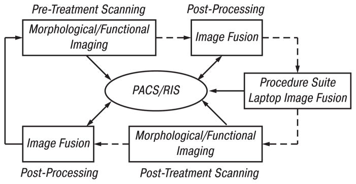
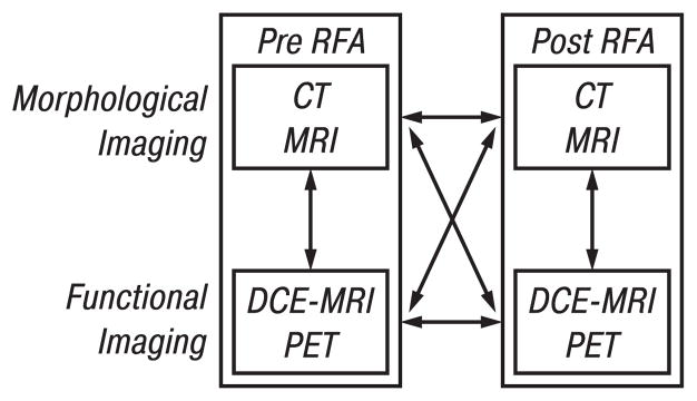
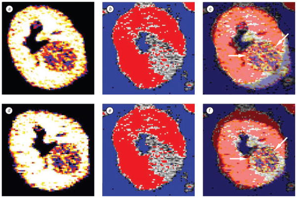
Methods: Multi-modality datasets were separately acquired that included functional (FDG-PET and DCE-MRI) and standard morphologic studies (CT and MRI). Different combinations of imaging modalities were registered and fused prior to, during, and following percutaneous image-guided tumor ablation with radiofrequency. Different algorithms and visualization tools were evaluated for both intra-modality and inter-modality image registration using the software MIPAV (Medical Image Processing, Analysis and Visualization). Semi-automated and automated registration algorithms were used on a standard PC workstation: 1) landmark-based least-squares rigid registration, 2) landmark-based thin-plate spline elastic registration, and 3) automatic voxel-similarity, affine registration.

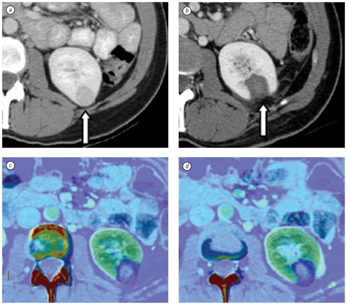
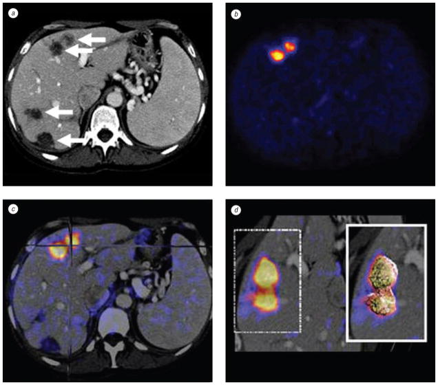
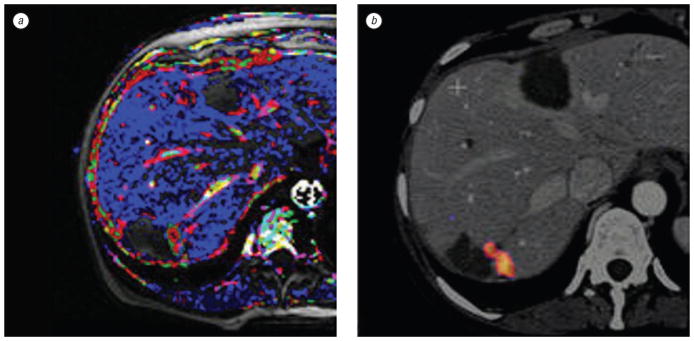
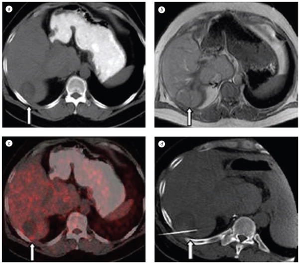
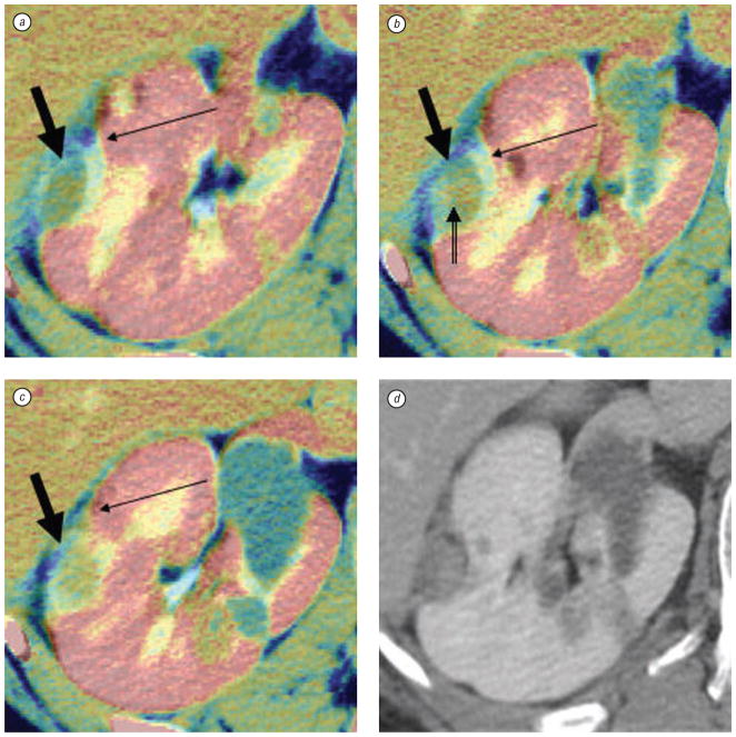
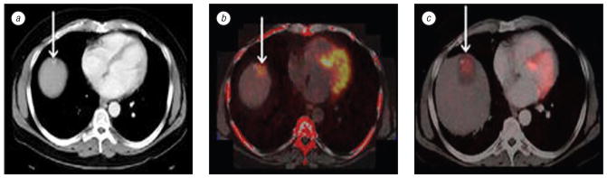
Results: Intra- and inter-modality image fusion were successfully performed prior to, during and after RFA procedures. Fusion of morphologic and functional images provided a useful view of the spatial relationship of lesion structure and functional significance. Fused axial images and segmented three-dimensional surface models were used for treatment planning and post-RFA evaluation, to assess potential for optimizing needle placement during procedures.

Conclusion: Fusion of morphologic and functional images is feasible before, during and after radiofrequency ablation of tumors in abdominal organs. For routine use, the semi-automated registration algorithms may be most practical. Image fusion may facilitate interventional procedures like RFA and should be further evaluated.


Figures

Fig. 1
Fig. 1
Workflow for patients scheduled for RFA. Initial imaging includes morphologic and/or functional imaging. DICOM images are sent directly or via PACS to a PC workstation for image registration and fusion. The results are pushed to a laptop that is taken into the procedure suite where patient information can be retrieved. During RFA, image fusion can be performed on the laptop, which may receive data from PACS or CT scanner. Follow-up morphologic and functional imaging data is later fused with pre-procedure imaging data on a workstation
Fig. 2
Fig. 2
Image fusion combines the visualization characteristics of malignant lesions seen with morphologic (CT, MRI) and functional (PET, DCE-MRI) imaging before, during, and after RFA. After RFA the relation and characteristics of tumor and thermal lesion can be assessed in a fused data set. Various combinations are possible, including intra-modality (e. g. pre-CT vs post-CT) or inter-modality (e. g. pre-CT vs pre-PET)
Fig. 3
Fig. 3
CT scan and elastic fusion images of kidney tumor and post-RFA thermal lesion. Contrast enhanced axial CT slices show a left kidney lesion before RFA (a, arrow) and thermal lesion two months after RFA (b, arrow). Due to change in kidney shape post ablation, elastic registration method is used to fuse pre- and post-RFA images (c and d), which defines treatment margins (dark blue)
Fig. 4
Fig. 4
Patient with metastatic pancreatic carcinoma isolated to the liver. Pre-RFA contrast enhanced axial CT slice (a) shows detailed morphology with 4 possible targets (arrows) for treatment. PET (b) shows abnormal FDG uptake in 2 anterior liver lesions. Intermodality PET/CT fusion (c) localizes active lesions (crosshairs). Volume rendering (d) visualizes metabolic activity with 3 dimensional details
Fig. 5
Fig. 5
Patient with residual tumor following RFA for multiple liver metastases. Dynamic contrast enhanced MRI (a) compared to a fused PET/CT (b) confirms tumor and correlates vascular pharmacokinetics with metabolic activity
Fig. 6
Fig. 6
CT, MRI, and PET images of a patient with colorectal carcinoma with liver metastases. Axial CT (a) and MRI (b) slices post-RFA showing only morphology (arrows) appearing negative for recurrence. Retrospective off-line fusion of CT and PET data sets (c) validates a pathology-proven residual tumor along the posterior border of the liver. Repeat treatment targeted with spatial knowledge of PET activity (d)
Fig. 7
Fig. 7
Right kidney tumor in patient with von Hipple-Lindau syndrome. Pre-RFA fused with post-RFA contrast enhanced axial CT slices (from superior to inferior, a–c) using optimized automated registration (OAR) method with correlation ratio voxel-similarity cost function. Images a–c were cropped and colorized with MIPAV tools to speed the registration process and enhance visualization. The pre-treatment CT appears in gray and the post-treatment CT is in color, i. e. the yellow treatment margin (thin arrows) overlays the original grayscale tumor (thick arrows). Notice the thin margin (double tailed arrow) in (b) which could potentially have been a site of recurrence; however, 6 month, 1 year, and 18 months (d) post treatment scans showed no recurrence. Although there was a thin margin, fusion correctly depicted an adequate thermal lesion
Fig. 8
Fig. 8
Patient with lesion at liver dome. Contrast enhanced axial CT slice shows low attenuation lesion adjacent to a high attenuation lesion (arrow) in dome of liver (a). Rigid registration of PET/CT pre-RFA shows abnormal FDG uptake over low attenuation lesion adjacent to high attenuation lesion (b). Post-RFA CT fused with pre-RFA PET (c) verifies RFA treatment zone with margins covering the area of abnormal FDG uptake (arrow)
Fig. 9
Fig. 9
Post processed cropped and colorized CT before RFA, after RFA, and fused image using rigid and elastic registration methods. Top row (a–c) is rigid registration whereas the bottom row (d–f) is elastic registration. The first column (a, d) is pre-RFA CT. The second column (b, e) is 2 months post RFA CT scan. Rigid registration (c) matches hand picked anatomy from one image to the other without altering either source image. This may result in mismatch (arrow) since the organ has shifted in the time interval between imaging. Elastic registration (f) also uses landmarks for point-to-point registration, but allows deformation of anatomy to better match the area of interest (arrow)

References

    1. Kramer EL, Noz ME, Sanger JJ, et al. CT-SPECT fusion to correlate radiolabeled monoclonal antibody uptake with abdominal CT findings. Radiology. 1989;172:861–5. - PubMed
    1. Pietrzyk U, Herholz K, Fink G, et al. An interactive technique for three-dimensional image registration: validation for PET, SPECT, MRI and CT brain studies. J Nucl Med. 1994;35:2011–8. - PubMed
    1. Wahl RL, Quint LE, Cieslak RD, et al. “Anatometabolic” tumor imaging: fusion of FDG PET with CT or MRI to localize foci of increased activity. J Nucl Med. 1993;34:1190–7. - PubMed
    1. Mountz JM, Zhang B, Liu HG, Inampudi C. A reference method for correlation of anatomic and functional brain images: validation and clinical application. Semin Nucl Med. 1994;24:256–71. - PubMed
    1. Correia JA. Registration of nuclear medicine images. J Nucl Med. 1990;31:1227–9. - PubMed

Publication types

LinkOut - more resources