

High temporal resolution OCT using image-based retrospective gating
- PMID: 19550478
- PMCID: PMC2748662
- DOI: 10.1364/oe.17.010786
High temporal resolution OCT using image-based retrospective gating
Abstract
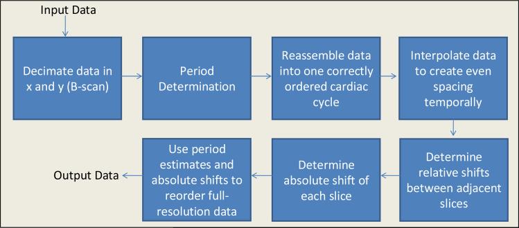
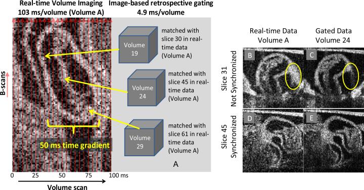
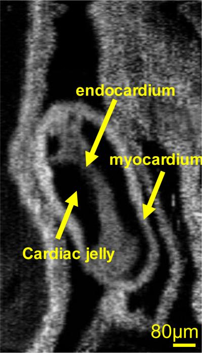
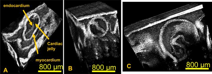
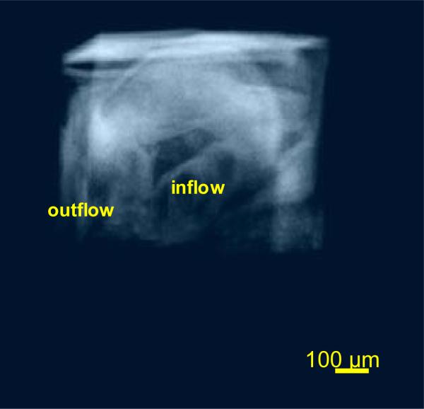
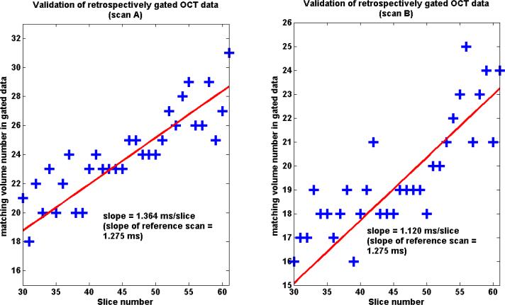
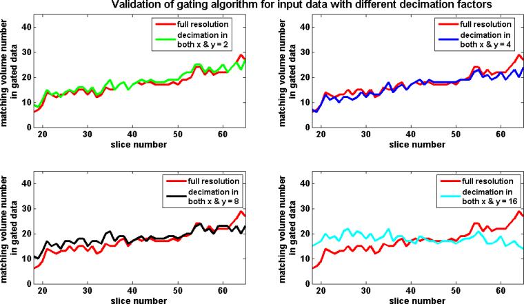
High temporal resolution OCT imaging is very advantageous for analyzing cardiac mechanics in the developing embryonic heart of small animals. An image-based retrospective gating technique is presented to increase the effective temporal resolution of an OCT system and to allow visualization of systolic dynamics in 3D. The gating technique employs image similarity measures for rearranging asynchronously acquired input data consisting of a time series of 2D images at each z position along the heart volume, to produce a time sequence of 3D volumes of the beating heart. The study includes a novel robust validation technique, which quantitatively evaluates the accuracy of the gating technique, in addition to visual evaluations by 2D multiplanar reformatting (MPR) and 3D volume rendering. The retrospective gating and validation is demonstrated on a stage 14 embryonic quail heart data set. Using the validation scheme, it is shown that the gating is accurate within a standard deviation of 4.7 ms, which is an order of magnitude shorter than the time interval during which systolic contraction (approximately 50 ms) occurs in the developing embryo. This gating method has allowed, for the first time, clear visualization of systolic dynamics of the looping embryonic heart in 3D.
Figures
Similar articles
-
4D reconstruction of the beating embryonic heart from two orthogonal sets of parallel optical coherence tomography slice-sequences.IEEE Trans Med Imaging. 2013 Mar;32(3):578-88. doi: 10.1109/TMI.2012.2231692. Epub 2012 Dec 4. IEEE Trans Med Imaging. 2013. PMID: 23221816 Free PMC article.
-
In vivo gated 4D imaging of the embryonic heart using optical coherence tomography.J Biomed Opt. 2007 May-Jun;12(3):030505. doi: 10.1117/1.2747208. J Biomed Opt. 2007. PMID: 17614708
-
Rotationally acquired four-dimensional optical coherence tomography of embryonic chick hearts using retrospective gating on the common central A-scan.J Biomed Opt. 2011 Sep;16(9):096007. doi: 10.1117/1.3622491. J Biomed Opt. 2011. PMID: 21950921 Free PMC article.
-
The diagnostic value of intracoronary optical coherence tomography.Herz. 2011 Aug;36(5):417-29. doi: 10.1007/s00059-011-3487-7. Herz. 2011. PMID: 21744151 Review.
-
Combining SLO and OCT technology.Bull Soc Belge Ophtalmol. 2006;(302):133-51. Bull Soc Belge Ophtalmol. 2006. PMID: 17265795 Review.
Cited by
-
Optical coherence tomography for embryonic imaging: a review.J Biomed Opt. 2016 May 1;21(5):50902. doi: 10.1117/1.JBO.21.5.050902. J Biomed Opt. 2016. PMID: 27228503 Free PMC article. Review.
-
Increasing the field-of-view of dynamic cardiac OCT via post-acquisition mosaicing without affecting frame-rate or spatial resolution.Biomed Opt Express. 2011 Sep 1;2(9):2614-22. doi: 10.1364/BOE.2.002614. Epub 2011 Aug 16. Biomed Opt Express. 2011. PMID: 22091446 Free PMC article.
-
Embryonic aortic arch hemodynamics are a functional biomarker for ethanol-induced congenital heart defects [Invited].Biomed Opt Express. 2017 Feb 24;8(3):1823-1837. doi: 10.1364/BOE.8.001823. eCollection 2017 Mar 1. Biomed Opt Express. 2017. PMID: 28663868 Free PMC article.
-
In vivo functional imaging of blood flow and wall strain rate in outflow tract of embryonic chick heart using ultrafast spectral domain optical coherence tomography.J Biomed Opt. 2012 Sep;17(9):96006-1. doi: 10.1117/1.JBO.17.9.096006. J Biomed Opt. 2012. PMID: 23085907 Free PMC article.
-
Motion-gated acquisition for in vivo optical imaging.J Biomed Opt. 2009 Nov-Dec;14(6):064038. doi: 10.1117/1.3275473. J Biomed Opt. 2009. PMID: 20059276 Free PMC article.
References
-
- Jenkins MW, Rothenberg F, Roy D, Nikolski VP, Hu Z, Watanabe M, Wilson DL, Efimov IR, Rollins AM. 4D embryonic cardiography using gated optical coherence tomography. Opt. Express. 2006;14:736–748. - PubMed
-
- Jenkins MW, Adler DC, Gargesha M, Huber R, Rothenberg F, Belding J, Watanabe M, Wilson DL, Fujimoto JG, Rollins AM. Ultrahigh-speed optical coherence tomography imaging and visualization of the embryonic avian heart using a buffered Fourier Domain Mode Locked laser. Opt. Express. 2007;15:6251–6267. - PMC - PubMed
-
- Jenkins MW, Chughtai OQ, Basavanhally AN, Watanabe M, Rollins AM. In vivo gated 4D imaging of the embryonic heart using optical coherence tomography. J. Biomed. Opt. 2007;12:030505. - PubMed
-
- Jenkins MW, Patel P, Deng H, Montano MM, Watanabe M, Rollins AM. Phenotyping transgenic embryonic murine hearts using optical coherence tomography. Appl. Opt. 2007;46:1776–1781. - PubMed
Publication types
MeSH terms
Grants and funding
LinkOut - more resources
Full Text Sources
