

Subcellular-resolution molecular imaging within living tissue by fiber microendoscopy
- PMID: 19550931
- PMCID: PMC3065245
- DOI: 10.1364/oe.15.016413
Subcellular-resolution molecular imaging within living tissue by fiber microendoscopy
Abstract
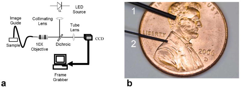
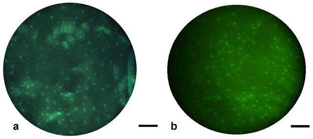
Conventional histopathology involves sampling, sectioning and staining of tissue specimens prior to microscopic evaluation, and provides diagnostic information at a single location and point in time. In vivo microscopy and molecular-targeted optical labeling are two rapidly developing fields, which together have the potential to provide anatomical and functional indications of disease by staining and imaging tissue in situ. To address the need for high-resolution imaging instrumentation, we have developed a compact, robust, and inexpensive fiber-optic microendoscopy system based around wide-field LED illumination, a flexible 1 mm diameter fiber-optic bundle, and a color CCD camera. We demonstrate the sub-cellular resolution imaging capabilities of the system through a series of experiments, beginning with simultaneous imaging of three different cancer cell lines in culture, each targeted with a distinct fluorescent label. We used the narrow diameter probe to access subcutaneous tumors in an in vivo murine model, allowing direct comparison of microendoscopy images with macroscopic images and histopathology. A surgically resected tissue specimen from the human oral cavity was imaged across the clinical margin, demonstrating qualitative and quantitative distinction between normal and cancerous tissue based on sub-cellular image features. Finally, the fiber-optic microendoscope was used on topically-stained normal human oral mucosa in vivo, resolving epithelial cell nuclei and membranes in real-time fluorescence images. Our results demonstrate that this imaging system can potentially complement conventional diagnostic techniques, and support efforts to translate emerging molecular-diagnostic and therapeutic agents into clinical use.
Figures
References
-
- Weissleder R, Tung CH, Mahmood U, Bogdanov A., Jr In vivo imaging of tumors with protease-activated near-infrared fluorescent probes. Nat Biotechnol. 1999;17:375–378. - PubMed
-
- Ke S, Xiaoxia W, Gurfinkel M, Charnsangavej C, Wallace S, Sevick-Muraca EM, Li C. Near-infrared optical imaging of epidermal growth factor receptor in breast cancer xenografts. Cancer Res. 2003;63:7870–7875. - PubMed
-
- Gao X, Cui Y, Levenson RM, Chung LWK, Nie S. In vivo cancer targeting and imaging with semiconductor quantum dots. Nat Biotechnol. 2004;22:969–976. - PubMed
-
- Becker A, Hessenius C, Licha K, Ebert B, Sukowski U, Semmler W, Wiedenmann B, Grötzinger C. Receptor-targeted optical imaging of tumors with near-infrared fluorescent ligands. Nat Biotechnol. 2001;19:327–331. - PubMed
-
- Jaffer FA, Weissleder R. Molecular imaging in the clinical arena. JAMA. 2005;293:855–862. - PubMed
Grants and funding
LinkOut - more resources
Full Text Sources
Other Literature Sources
Medical
