. 2008 Dec 1;80(23):9298-309.
doi: 10.1021/ac801654h.

Multiple isotopic labels for quantitative mass spectrometry

Affiliations

Multiple isotopic labels for quantitative mass spectrometry

Cain Morano et al. Anal Chem. .

Abstract

Quantitative mass spectrometry is often performed using isotopically labeled samples. Although the 4-trimethylammoniumbutyryl (TMAB) labels have many advantages over other isotopic tags, only two forms have previously been synthesized (i.e., a heavy form containing nine deuteriums and a light form without deuterium). In the present report, two additional forms containing three and six deuteriums have been synthesized and tested. These additional isotopic tags perform identically to the previously reported tags; peptides labeled with the new TMAB reagents coelute from reversed-phase HPLC columns with peptides labeled with the lighter and heavier TMAB reagents. Altogether, these four tags allow for multivariate analysis in a single liquid chromatography/mass spectrometry analysis, with each isotopically tagged peptide differing in mass by 3 Da per tag incorporated. The synthetic scheme is described in simple terms so that a biochemist without specific training in organic chemistry can perform the synthesis. The interpretation of tandem mass spectrometry data for the TMAB-labeled peptides is also described in more detail. The additional TMAB isotopic reagents described here, together with the additional description of the synthesis and analysis, should allow these labels to be more widely used for proteomics and peptidomics analyses.


Figures

Figure 1
Figure 1
Synthetic scheme of TMAB labels. Step (a) is the alkylation of gamma-amino butyric acid (GABA) using methyl iodide or one of its deuterated isotopes. This generates a trimethylammonium iodide salt, which is washed with chloroform, and then converted to the chloride salt by acidification with HCl. Step (b) is the activation of the acid by formation of the N-hydroxysuccinimide (NHS) ester using dicyclohexylcarbodiimide (DCC).
Figure 2
Figure 2
Chemical structures of the TMAB labels. The net masses, in Daltons, are of the carbon chain, the carbonyl oxygen, and the trimethyl ammonium group, minus the N-hydroxy succinimide group and the chloride ion (i.e. the mass difference in a peptide after incorporating one of these compounds on a free N-terminal amine).
Figure 3
Figure 3
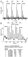
MS spectra of peptides labeled with the isotopic TMAB tags and analyzed by LC/MS. A: Example of a peptide that displays baseline resolution between each isotopic tag. The peptide was identified by MS/MS analysis as neuromedin N (KIPYIL) which incorporated 2 isotopic tags. The indicated monoisotopic m/z values represent the 2+ ion. B: Example of a peptide with relatively smaller mass differences between isotopic peaks and which therefore shows substantial peak overlap. This peptide was identified by MS/MS analysis as a chromogranin B-derived peptide corresponding to residues 438–454 (LLDEGHYPVRESPIDTA). The indicated monoisotopic m/z values represent the 3+ ion that incorporated 1 isotopic tag (on the N-terminus) and protons on the His and Arg residues. To determine the relative abundance of each isotopic form, it is necessary to subtract the contribution from the 13C-containing peptides of the lower m/z values. For this, Protein Prospector was used to determine the natural isotope distribution of this peptide and the baseline of each peak was then adjusted to account for the contribution from the 13C-containing isotope of the lower m/z peak, as shown in panel B.
Figure 4
Figure 4
The 4 isotopically-labeled forms of each peptide co-elute from a reverse phase LC column. Left panels represent chromogranin B (64–86) and the right panels represent chromogranin B (438–454). The relative intensity for m/z values representing each isotopic form are plotted versus elute time in minutes.
Figure 5
Figure 5
Representative MS/MS spectra. (A) MS/MS of the proenkephalin-derived peptide SPQLEDEAKELQ, which incorporated two D0-TMAB tags. Note the complete loss of trimethylamine from the observed a, b, and y series ions. The spectrum also shows an MH2+ peak for the intact peptide, and stronger peaks corresponding to the loss of 59 and 118 Da (i.e. 2×59); these represent neutral loss of one and two trimethylamines from the parent ion. (B) MS/MS of D9-TMAB-labeled proSAAS-derived peptide SLSAASAPLVETSTPL. For this peptide, many of the trimethylamine groups are not lost in the collision-induced-dissociation, so most of the b series ions are indicated as +68 (for D9-TMAB-labeled peptide). In this example, partial breakdown of tags leads to inconclusive results upon Mascot database searches, so manual interpretation is required.
Figure 6
Figure 6
Distribution of peak ratios of the 107 peptides detected in the analysis of mouse brain. The peak-to-peak ratios of each peptide fragment were measured and the individual peak intensity for each isotopically-labeled form was compared to the average peak intensity of the other 3 isotopically-labeled forms of the peptide (see Table 1; columns 8–11). Then, the number of peptides for each ratio was plotted versus the observed ratio. The theoretical ratio is 1.00, and nearly all of the isotopically-labeled forms of each peptide showed ratios within 20% of this theoretical ratio.

References

    1. Strand FL. Prog Drug Res. 2003;61:1–37. - PubMed
    1. Hokfelt T, Broberger C, Xu ZQ, Sergeyev V, Ubink R, Diez M. Neuropharm. 2000;39:1337–56. - PubMed
    1. Hummon AB, Amare A, Sweedler JV. Mass Spectrom Rev. 2006;25:77–98. - PubMed
    1. Hummon AB, Richmond TA, Verleyen P, Baggerman G, Huybrechts J, Ewing MA, Vierstraete E, Rodriguez-Zas SL, Schoofs L, Robinson GE, Sweedler JV. Science. 2006;314:647–49. - PubMed
    1. Yuan X, Desiderio DM. J Mass Spectrom. 2005;40:176–81. - PubMed

Publication types