. 2009 Aug;42(4):413-24.
doi: 10.1111/j.1365-2184.2009.00612.x. Epub 2009 Jun 23.

Transmitochondrial embryonic stem cells containing pathogenic mtDNA mutations are compromised in neuronal differentiation

Affiliations

Transmitochondrial embryonic stem cells containing pathogenic mtDNA mutations are compromised in neuronal differentiation

D M Kirby et al. Cell Prolif. 2009 Aug.

Abstract

Objectives: Defects of the mitochondrial genome (mtDNA) cause a series of rare, mainly neurological disorders. In addition, they have been implicated in more common forms of movement disorders, dementia and the ageing process. In order to try to model neuronal dysfunction associated with mitochondrial disease, we have attempted to establish a series of transmitochondrial mouse embryonic stem cells harbouring pathogenic mtDNA mutations.

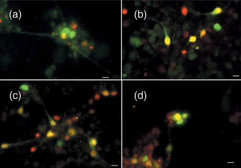
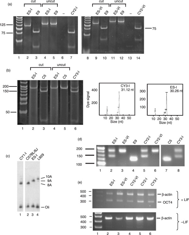
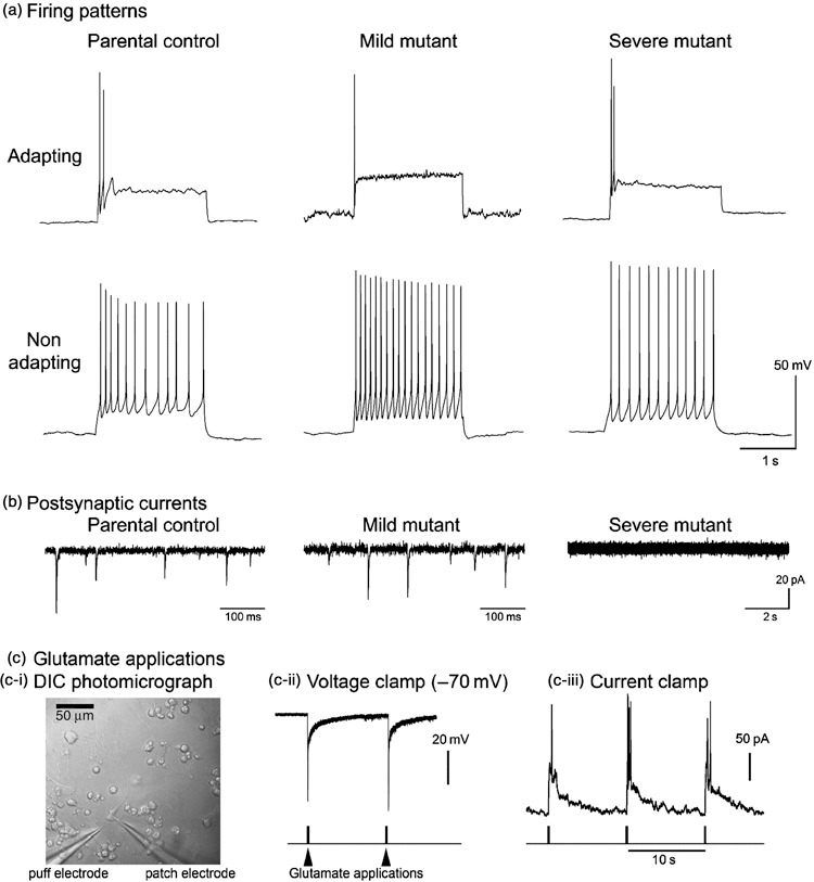
Materials and methods: Transmitochondrial embryonic stem cell cybrids were generated by fusion of cytoplasts carrying a variety of mtDNA mutations, into embryonic stem cells that had been pretreated with rhodamine 6G, to prevent transmission of endogenous mtDNA. Cybrids were differentiated into neurons and assessed for efficiency of differentiation and electrophysiological function.

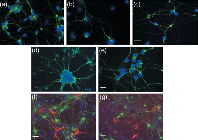
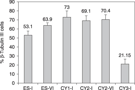
Results: Neuronal differentiation could occur, as indicated by expression of neuronal markers. Differentiation was impaired in embryonic stem cells carrying mtDNA mutations that caused severe biochemical deficiency. Electrophysiological tests showed evidence of synaptic activity in differentiated neurons carrying non-pathogenic mtDNA mutations or in those that caused a mild defect of respiratory activity. Again, however, neurons carrying mtDNA mutations that resulted in severe biochemical deficiency had marked reduction in post-synaptic events.

Conclusions: Differentiated neurons carrying severely pathogenic mtDNA defects can provide a useful model for understanding how such mutations can cause neuronal dysfunction.


Figures

Figure 3
Figure 3
GABAergic neurons are present in subpopulations of differentiated transmitochondrial cells. Following 15–16 days of differentiation, cells were analysed for the expression of an early neuronal marker (NeuN, red) and the presence of GABA (green) as detailed in the Materials and methods. Co‐expression is shown in the image as yellow. The size marker represents 10 µm. (a) Differentiated parental ES‐I; (b) CY1‐I carrying the polymorphic marker; (c) CY2‐I carrying the mild respiratory defect; (d) CY3‐I carrying the severe respiratory defect.
Figure 1
Figure 1
Production of transmitocondrial murine embryonic stem (ES) cell cybrids. (a) Transfer of mtDNA carrying a mild mutation in Mtco1 to pluripotent ES‐I and ES‐VI ES cell lines. A 133‐bp amplicon spanning nt6589 was generated from the indicated ES parental cells (ES‐I, lanes 2 and 4; ES‐VI, lanes 9 and 11), 6589T>C fibroblasts (E9, lanes 3, 5, 10 and 12) and ES cybrids (CY2‐I, lanes 7 and 14) as detailed in the Materials and methods and subjected to digestion with Cfo1. Cleavage generated a 115‐ and 18‐bp fragment for the wild‐type amplicon (lanes 2, 9), with further digestion of the 115‐bp fragment to 75 and 40 bp indicating the 6589T>C mutation (lanes 3, 7, 10 and 14). Uncut amplicon (lanes 4, 5, 11 and 12), negative control (lanes 6 and 13) and molecular weight markers (lanes 1, 8) are also shown. (b) Transfer of mutations in Mtnd5 and Mtnd6 to ES‐I. A 182‐bp amplicon spanning nt12273 was generated from ES parental cells (ES‐I, lanes 2 and 4), 12273G>A fibroblasts (C5, lanes 3 and 5) and ES cell cybrids (CY3‐I, lane 6) as detailed in the Materials and methods and subjected to cleavage with Dde1. Products carrying the mutation were cleaved to generate fragments of 145 and 37 bp (lanes 3 and 6). Uncut amplicon (lanes 4 and 5) and molecular weight ladder (lane 1) are shown. For analysis of mtDNA carrying the 13887C insertion, primer extension was performed as detailed in the Materials and methods. The panels show the length of extension corresponding to the normal mtDNA sequence (ES‐I, ~30 bp) or to the mtDNA carrying the single base insertion (CY3‐I, ~31 bp). The fluorescence signal due to the 5′ D3 fluorescent conjugate is indicated. The conjugated standard (13 nt) is also shown. (c) Transfer of the Mttr polymorphism to ES‐I. Primer extension was used to determine the length of the polyA tract around nt9821 in the mtDNA of various cell lines as detailed in the Materials and methods. Parental ES cells (ES‐I, lane 3), donor fibroblasts (C57BL/6J, lane 2) and cybrids (CY1‐I, lane 1) are shown, as well as the 10A extension found in L929 fibroblasts (lane 4). Radiolabelled oligonucleotide is also shown (oli). (d) Microsatellite markers confirm production of transmitochondrial cybrids. Amplicons spanning the informative marker D6mit102 were prepared from ES parental cells (ES‐I, lane 2; ES‐VI, lane 3), fibroblast donors (E9, lane 4; C5, lane 7) and cybrids (CY2‐I, lane 5; CY2‐VI, lane 6; CY3‐I, lane 8). Molecular weight markers are shown in lane 1. (e) Oct‐4 expression in parental and cybrid ES cells is dependent on leukaemia inhibitory factor (LIF). Cell lines (ES parental lanes 2, 3; cybrids lanes 4–6) were grown in the presence (+) or absence (–) of LIF for 9 days before RNA was isolated and reverse transcribed. Amplicons from the transcripts encoding β‐actin or Oct‐4 were generated as described in the Materials and methods. Molecular weight standards are given (lane 1).
Figure 2
Figure 2
Transmitochondrial embryonic stem (ES) cells can be differentiated into neurons and astrocytes. Parental ES cell lines (a and f, ES‐I; d, ES‐VI) and cybrids (b and g, CY3‐I SEVERE; c, CY2‐I MILD; e, CY2‐VI MILD) were differentiated and fixed on Day 13 for immunohistochemical analysis with the pan‐neuronal anti‐β‐tubulin III antibody (a–g, green) and astrocytic marker anti‐GFAP (F and G, red) as detailed in the Materials and methods. Nuclei are visualized by DAPI staining (blue). The sizing bar represents 20 µm.
Figure 4
Figure 4
Severe respiratory deficiency affects neuronal differentiation of murine embryonic stem (ES) cells. Cell lines were induced to differentiate as described in the Materials and methods. On day 13, cells were subjected to immunohistochemistry with anti‐β‐tubulin III (late pan‐neuronal marker). Fields (n = 10) were selected and the percentage of cells differentiating into neurons was calculated with reference to the total number of DAPI‐staining nuclei in the field. This was done for two independent differentiation experiments, i.e. the results represent 2 × 10 fields at ×100 magnification, for all except for CY1‐I, in which efficiency of differentiation into neurons was quantified only once (1 × 10 fields). ES‐I and ES‐VI, parental murine ES cells; CY1‐I, polymorphism; CY3‐I, severe respiratory defect; CY2‐I and CY2‐VI, mild respiratory defect.
Figure 5
Figure 5
All cell lines produce neurons with a range of firing patterns. (a) Example of adapting and non‐adapting firing patterns are shown in response to square pulses of current injected somatically. Individual cells only ever show one firing pattern, but examples of each type are found in all cell lines. (b) Typical examples of spontaneous synaptic currents recorded in cultured neurons held at –70 mV. In the control and mild mitochondrial mutant cell lines, synaptic currents are routinely recorded in all cells after about the 2nd week after plating (parental recording is at 6 days post‐plating; mild mutant recording – 13 days post‐plating). In contrast, recordings from the severe mutant cell line are routinely devoid of synaptic currents at all ages recorded (example is at post‐plating 17). (c) Glutamate applications. Panel (c‐i) shows a differential interference micrograph of the experimental arrangement with the puff electrode located 10–20 µm from the patched cell. Panels (c‐ii) and (c‐iii) show responses to exogenously applied glutamate (1 mm) in a cell from the severe mutant line that showed no spontaneous synaptic events in more than 5 min of recording.

References

    1. Taylor RW, Turnbull DM (2005) Mitochondrial DNA mutations in human disease. Nat. Rev. Genet. 6, 389–402. - PMC - PubMed
    1. Bender A, Krishnan KJ, Morris CM, Taylor GA, Reeve AK, Perry RH et al (2006) High levels of mitochondrial DNA deletions in substantia nigra neurons in aging and Parkinson disease. Nat. Genet. 38, 515–517. - PubMed
    1. Trifunovic A, Wredenberg A, Falkenberg M, Spelbrink JN, Rovio AT, Bruder CE et al (2004) Premature ageing in mice expressing defective mitochondrial DNA polymerase. Nature 429, 417–423. - PubMed
    1. Vermulst M, Wanagat J, Kujoth GC, Bielas JH, Rabinovitch PS, Prolla TA et al (2008) DNA deletions and clonal mutations drive premature aging in mitochondrial mutator mice. Nat. Genet. 40, 392–394. - PubMed
    1. Fan W, Waymire KG, Narula N, Li P, Rocher C, Coskun PE et al (2008) A mouse model of mitochondrial disease reveals germline selection against severe mtDNA mutations. Science 319, 958–962. - PMC - PubMed

Publication types

MeSH terms

Substances