

Structures involved at the time of temporal lobe spikes revealed by interindividual group analysis of EEG/fMRI data
- PMID: 19552652
- PMCID: PMC3769286
- DOI: 10.1111/j.1528-1167.2009.02180.x
Structures involved at the time of temporal lobe spikes revealed by interindividual group analysis of EEG/fMRI data
Abstract
Purpose: We measured metabolic changes associated with temporal lobe (TL) spikes using combined electroencephalography (EEG) and functional magnetic resonance imaging (fMRI). We selected 18 patients with temporal lobe epilepsy (TLE) who underwent a 2-h simultaneous EEG-fMRI and had unilateral or bilateral independent TL spikes for interindividual group analysis, in order to identify consistent blood oxygenation level dependent (BOLD) responses to TL spikes.
Methods: EEG was postprocessed and spikes were visually identified. fMRI data were preprocessed with motion correction, spatial smoothing, and removal of low frequency drifts. Spike timings were used as events for fMRI statistical analysis. Four hemodynamic response functions were used to account for variability in the BOLD response.
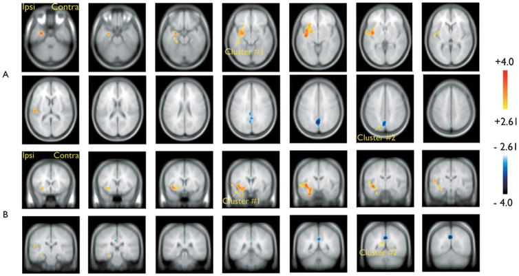
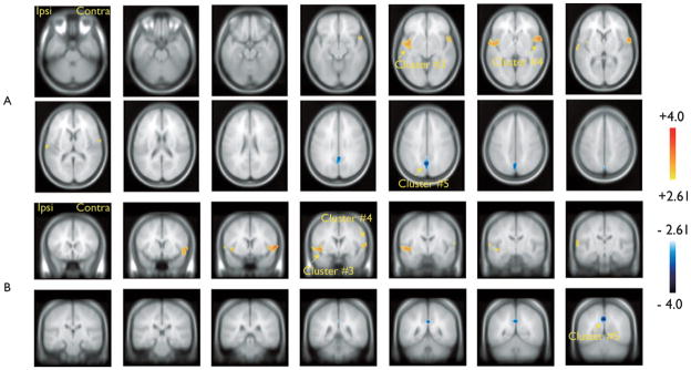
Results: Group analysis revealed common areas of BOLD activations and deactivations. The hemodynamic response function (HRF) peaking 3 s after the spike showed activation involving ipsilaterally the mesial temporal structures (presumably the hippocampus), putamen/globus pallidus, inferior insula, and superior temporal gyrus. The HRF peaking at 5 s showed activations involving ipsi- and contralaterally the superior temporal gyrus and inferior insula. Both HRFs showed bilateral posterior cingulate deactivations.
Discussion: We disclosed involvement of a network of activated areas during unilateral TL spikes, including ipsilateral mesial temporal structures, basal ganglia, and bilateral neocortical temporal regions. Despite the low temporal resolution of fMRI we demonstrated that contralateral temporal involvement occurred later than ipsilateral activation. This contralateral change took place in the absence of visible EEG changes. The posterior cingulate deactivation may reflect the interconnections between this region and other limbic structures. It may also partially correspond to a suspension of the default mode network, as previously described for TL spikes.
Conflict of interest statement
Disclosure: None of the authors has any conflict of interest to disclose.
Figures
Similar articles
-
Temporal and extratemporal BOLD responses to temporal lobe interictal spikes.Epilepsia. 2006 Feb;47(2):343-54. doi: 10.1111/j.1528-1167.2006.00427.x. Epilepsia. 2006. PMID: 16499759
-
EEG spectral changes underlying BOLD responses contralateral to spikes in patients with focal epilepsy.Epilepsia. 2009 Jul;50(7):1804-9. doi: 10.1111/j.1528-1167.2009.02080.x. Epub 2009 Apr 6. Epilepsia. 2009. PMID: 19389143
-
Temporal lobe spikes: EEG-fMRI contributions to the "mesial vs. lateral" debate.Clin Neurophysiol. 2017 Jun;128(6):986-991. doi: 10.1016/j.clinph.2017.03.041. Epub 2017 Apr 9. Clin Neurophysiol. 2017. PMID: 28445839
-
Epileptic networks studied with EEG-fMRI.Epilepsia. 2008;49 Suppl 3(Suppl 3):42-51. doi: 10.1111/j.1528-1167.2008.01509.x. Epilepsia. 2008. PMID: 18304255 Free PMC article. Review.
-
Temporal lobe epilepsy: when are invasive recordings needed?Epilepsia. 2000;41 Suppl 3:S61-74. doi: 10.1111/j.1528-1157.2000.tb01536.x. Epilepsia. 2000. PMID: 11001338 Review.
Cited by
-
Study on the Relationships between Intrinsic Functional Connectivity of the Default Mode Network and Transient Epileptic Activity.Front Neurol. 2014 Oct 10;5:201. doi: 10.3389/fneur.2014.00201. eCollection 2014. Front Neurol. 2014. PMID: 25346721 Free PMC article.
-
Temporal lobe epilepsy and affective disorders: the role of the subgenual anterior cingulate cortex.J Neurol Neurosurg Psychiatry. 2015 Feb;86(2):144-51. doi: 10.1136/jnnp-2013-306966. Epub 2014 May 29. J Neurol Neurosurg Psychiatry. 2015. PMID: 24876189 Free PMC article.
-
Identification of pre-spike network in patients with mesial temporal lobe epilepsy.Front Neurol. 2014 Oct 28;5:222. doi: 10.3389/fneur.2014.00222. eCollection 2014. Front Neurol. 2014. PMID: 25389415 Free PMC article.
-
Temporal Lobe Spikes Affect Distant Intrinsic Connectivity Networks.Front Neurol. 2021 Dec 17;12:746468. doi: 10.3389/fneur.2021.746468. eCollection 2021. Front Neurol. 2021. PMID: 34975714 Free PMC article.
-
Electrophysiological correlates of the BOLD signal for EEG-informed fMRI.Hum Brain Mapp. 2015 Jan;36(1):391-414. doi: 10.1002/hbm.22623. Epub 2014 Oct 3. Hum Brain Mapp. 2015. PMID: 25277370 Free PMC article. Review.
References
-
- Aarts JHP, Binnie CD, Smit AM, Wilkins AJ. Selective cognitive impairment during focal and generalized epileptiform EEG activity. Brain. 1984;107:293–308. - PubMed
-
- Archer JS, Abbott DF, Waites AB, Jackson GD. fMRI ‘deactivation’ of the posterior cingulate during generalized spike and wave. Neuroimage. 2003;20:1915–1922. - PubMed
-
- Blumenfeld H, McNally KA, Vanderhill SD, Paige AL, Chung R, Davis K, Norden AD, Stokking R, Studholme C, Novotny EJ, Jr, Zubal IG, Spencer SS. Positive and negative network correlations in temporal lobe epilepsy. Cereb Cortex. 2004;14:892–902. - PubMed
-
- Bonilha L, Rorden C, Castellano G, Pereira F, Rio PA, Cendes F, Li LM. Voxel-based morphometry reveals gray matter network atrophy in refractory medial temporal lobe epilepsy. Arch Neurol. 2004;61:1379–1384. - PubMed
Publication types
MeSH terms
Substances
Grants and funding
LinkOut - more resources
Full Text Sources
Medical
