

Distributed source modeling of language with magnetoencephalography: application to patients with intractable epilepsy
- PMID: 19552656
- PMCID: PMC2760088
- DOI: 10.1111/j.1528-1167.2009.02172.x
Distributed source modeling of language with magnetoencephalography: application to patients with intractable epilepsy
Abstract
Purpose: To examine distributed patterns of language processing in healthy controls and patients with epilepsy using magnetoencephalography (MEG), and to evaluate the concordance between laterality of distributed MEG sources and language laterality as determined by the intracarotid amobarbital procedure (IAP).
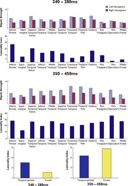
Methods: MEG was performed in 10 healthy controls using an anatomically constrained, noise-normalized distributed source solution (dynamic statistical parametric map, dSPM). Distributed source modeling of language was then applied to eight patients with intractable epilepsy. Average source strengths within temporoparietal and frontal lobe regions of interest (ROIs) were calculated, and the laterality of activity within ROIs during discrete time windows was compared to results from the IAP.
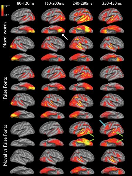
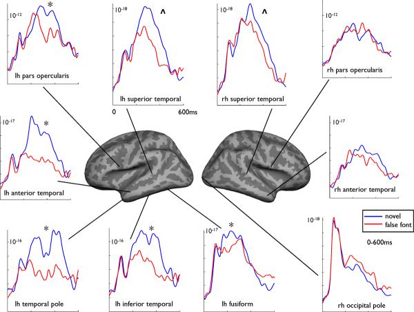
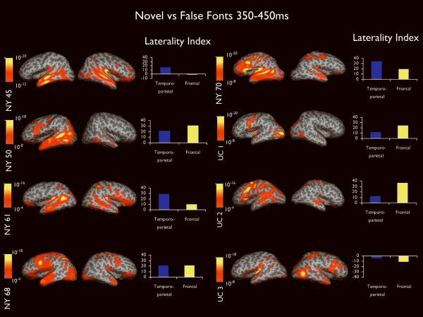
Results: In healthy controls, dSPM revealed activity in visual cortex bilaterally from approximately 80 to 120 ms in response to novel words and sensory control stimuli (i.e., false fonts). Activity then spread to fusiform cortex approximately 160-200 ms, and was dominated by left hemisphere activity in response to novel words. From approximately 240 to 450 ms, novel words produced activity that was left-lateralized in frontal and temporal lobe regions, including anterior and inferior temporal, temporal pole, and pars opercularis, as well as bilaterally in posterior superior temporal cortex. Analysis of patient data with dSPM demonstrated that from 350 to 450 ms, laterality of temporoparietal sources agreed with the IAP 75% of the time, whereas laterality of frontal MEG sources agreed with the IAP in all eight patients.
Discussion: Our results reveal that dSPM can unveil the timing and spatial extent of language processes in patients with epilepsy and may enhance knowledge of language lateralization and localization for use in preoperative planning.
Figures
References
-
- ALLISON T, PUCE A, SPENCER DD, MCCARTHY G. Electrophysiological studies of human face perception. I: Potentials generated in occipitotemporal cortex by face and non-face stimuli. Cereb Cortex. 1999;9:415–30. - PubMed
-
- BILLINGSLEY-MARSHALL RL, CLEAR T, MENCL WE, SIMOS PG, SWANK PR, MEN D, SARKARI S, CASTILLO EM, PAPANICOLAOU AC. A comparison of functional MRI and magnetoencephalography for receptive language mapping. J Neurosci Methods. 2007;161:306–13. - PubMed
-
- BOWYER SM, FLEMING T, GREENWALD ML, MORAN JE, MASON KM, WEILAND BJ, SMITH BJ, BARKLEY GL, TEPLEY N. Magnetoencephalographic localization of the basal temporal language area. Epilepsy Behav. 2005a;6:229–34. - PubMed
-
- BOWYER SM, MORAN JE, WEILAND BJ, MASON KM, GREENWALD ML, SMITH BJ, BARKLEY GL, TEPLEY N. Language laterality determined by MEG mapping with MR-FOCUSS. Epilepsy Behav. 2005b;6:235–41. - PubMed
-
- BREIER JI, CASTILLO EM, SIMOS PG, BILLINGSLEY-MARSHALL RL, PATARAIA E, SARKARI S, WHELESS JW, PAPANICOLAOU AC. Atypical language representation in patients with chronic seizure disorder and achievement deficits with magnetoencephalography. Epilepsia. 2005;46:540–8. - PubMed
Publication types
MeSH terms
Substances
Grants and funding
LinkOut - more resources
Full Text Sources
Medical
Research Materials
Miscellaneous
