. 2009 Jun 24:9:200.
doi: 10.1186/1471-2407-9-200.

Production and characterisation of monoclonal antibodies against RAI3 and its expression in human breast cancer

Affiliations

Production and characterisation of monoclonal antibodies against RAI3 and its expression in human breast cancer

Hannah Jörissen et al. BMC Cancer. .

Abstract

Background: RAI3 is an orphan G-protein coupled receptor (GPCR) that has been associated with malignancy and may play a role in the proliferation of breast cancer cells. Although its exact function in normal and malignant cells remains unclear and evidence supporting its role in oncogenesis is controversial, its abundant expression on the surface of cancer cells would make it an interesting target for the development of antibody-based therapeutics. To investigate the link with cancer and provide more evidence for its role, we carried out a systematic analysis of RAI3 expression in a large set of human breast cancer specimens.

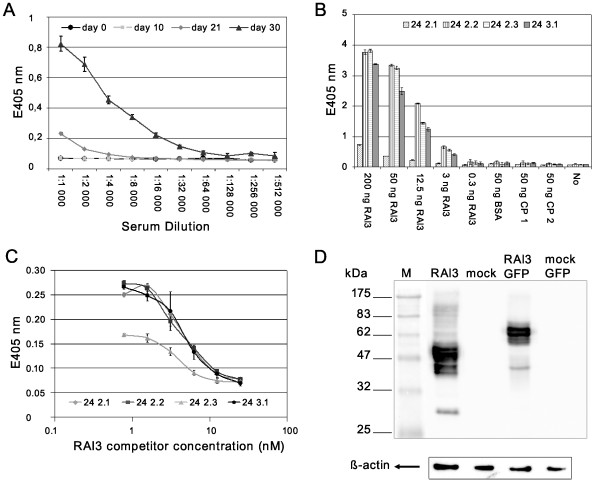
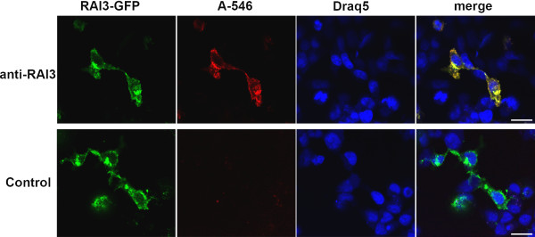
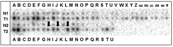
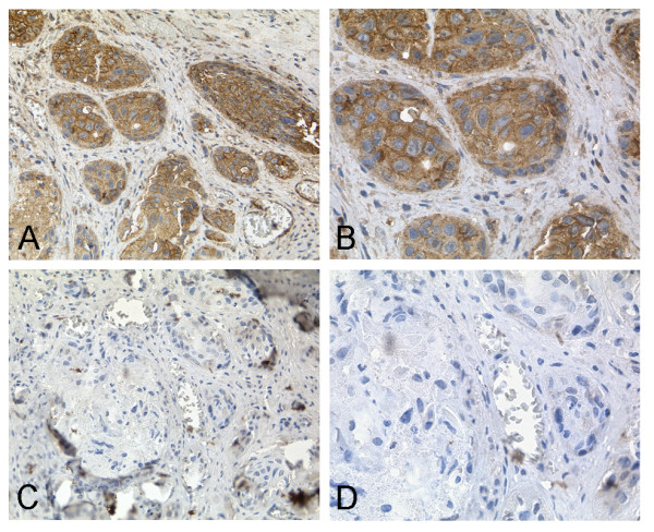
Methods: We expressed recombinant human RAI3 in bacteria and reconstituted the purified protein in liposomes to raise monoclonal antibodies using classical hybridoma techniques. The specific binding activity of the antibodies was confirmed by enzyme-linked immunosorbent assay (ELISA), western blot and immunocytochemistry. We carried out a systematic immunohistochemical analysis of RAI3 expression in human invasive breast carcinomas (n = 147) and normal breast tissues (n = 44) using a tissue microarray. In addition, a cDNA dot blot hybridisation assay was used to investigate a set of matched normal and cancerous breast tissue specimens (n = 50) as well as lymph node metastases (n = 3) for RAI3 mRNA expression.

Results: The anti-RAI3 monoclonal antibodies bound to recombinant human RAI3 protein with high specificity and affinity, as shown by ELISA, western blot and ICC. The cDNA dot blot and immunohistochemical experiments showed that both RAI3 mRNA and RAI3 protein were abundantly expressed in human breast carcinoma. However, there was no association between RAI3 protein expression and prognosis based on overall and recurrence-free survival.

Conclusion: We have generated a novel, highly-specific monoclonal antibody that detects RAI3 in formaldehyde-fixed paraffin-embedded tissue. This is the first study to report a systematic analysis of RAI3 expression in normal and cancerous human breast tissue at both the mRNA and protein levels.


Figures

Figure 1
Figure 1
Characterisation of monoclonal anti-RAI3 antibodies. A:. Direct ELISA of a serial dilution of BALB/c mouse serum carried out at several time points during immunisation. Increasing antbody titre indicates a good humoral immune response. Measurements are from triplicates, expressed as means ± S.D. B: Direct ELISA of hybridoma supernatants from four selected monoclones showing a concentration dependent RAI3 signal and no cross-reactivity to control proteins. BSA: bovine serum albumin; CP1: control protein 1, non-related human protein (CD30 ligand); CP2: control protein 2, related human protein, GPCR (GPR30); No: Negative control, no protein coated. Measurements are from triplicates, expressed as means ± S.D. C: Competitive ELISA of Mabs pre-incubated with a serial dilution of soluble antigen RAI3. Only unbound antibody can bind to the immobilised antigen on the ELSA plate allowing the comparison and determination of affinities based on the absorption values. Measurements are from triplicates, expressed as means ± S.D. D: Western blot analysis of cell extracts from transfected HEK293T cells. Cells expressed either RAI3 (lane 1), mock protein (lane 2), RAI3-GFP (lane 3) or mock-GFP protein (lane 4). Aliquots (10 μg) of total protein from cell extracts are loaded as indicated, with β-actin as the loading control. At least three experiments for Mab 24 2.3 are represented. Equal binding pattern was observed in all experiments.
Figure 2
Figure 2
Immunocytochemistry analysis of HEK293T cells expressing RAI3-GFP. Transiently transfected HEK293T cells expressing RAI3-GFP were subjected to immunocytochemistry analysis. The upper panels (anti-RAI3) show transfected cells incubated with anti-RAI3 antibody and subsequently with anti mouse AlexaFluor 546-labelled secondary antibody (A546). The immunostaning is pseudo-coloured in red. Co-localisation of the RAI3-GFP and A546 signals is shown in yellow in the merge panel. For the transfected control cells (Control panels) primary anti-RAI3 antibody was omitted in order to exclude unspecific binding of the A546 secondary antibody. Nuclei were stained using Draq5 and are pseudo-coloured in blue. Imaging was carried out by confocal microscopy using a 40× air objective. Scale bar: 20 μm
Figure 3
Figure 3
cDNA dot blot analysis of RAI3 expression in matched normal and t breast tissue. Expression profiles were determined using cDNA dot blot hybridisation analysis (BD Clontech) containing cDNA pairs derived from 50 matched normal breast tissues (N), tumourous breast tissues (T), and three lymph node metastastic tissues (marked with arrows). Upregulation of RAI3 was observed in 30 of 50 primary breast tumours as well as in one of three metastatic lymph nodes, as compared to matched normal breast tissue.
Figure 4
Figure 4
RAI3 Immunohistochemistry on TMA derived from normal and cancerous breast tissue. Immunohistochemical expression analysis of RAI3 in normal breast tissue as well as non-invasive and invasive breast tumours using a tissue microarray. (A, B) In normal breast epithelial cells RAI3 expression (IRS = 8) was often weaker than in invasive breast carcinoma cells. (C, D) In ductal carcinoma in situ RAI3 expression (IRS = 3) was often less intense compared to invasive breast carcinoma. (E, F) In invasive breast carcinomas (here: ductal type) RAI3 was often expressed more abundantly in the cytoplasm and in the cell membrane (example shows staining with an IRS = 12) than in either ductal carcinoma in situ or normal breast tissue. (G, H) In tubular breast carcinomas, RAI3 expression was less abundant (IRS = 3) than in most invasive ductal breast carcinomas. (I, J) In mucinous breast carcinomas, RAI3 expression was also less intense (IRS = 1) in comparison to most invasive ductal breast cancer cells. Magnifications: A, C, E, G, I: 40×; B, D, F, H, J: 400×.
Figure 5
Figure 5
Competitive RAI3 Immunohistochemistry on cancerous breast tissue. Immunohistochemical staining of cancerous breast tissue (A, B) in comparison to a competitive immunohistochemical approach (C, D). Strong staining of tumourous tissue was observed using a 1:50 dilution of anti-RAI3 Mab 24 2.3 (A, B). Staining is inhibited by pre-incubation of the antibody with 200-fold molar excess of recombinant RAI3 protein (C, D). Magnifications: A, C: 200×; B, D: 400×.

References

    1. Li S, Huang S, Peng SB. Overexpression of G protein-coupled receptors in cancer cells: involvement in tumor progression. Int J Oncol. 2005;27(5):1329–1339. - PubMed
    1. McCudden CR, Hains MD, Kimple RJ, Siderovski DP, Willard FS. G-protein signaling: back to the future. Cell Mol Life Sci. 2005;62(5):551–577. doi: 10.1007/s00018-004-4462-3. - DOI - PMC - PubMed
    1. Cheng Y, Lotan R. Molecular cloning and characterization of a novel retinoic acid-inducible gene that encodes a putative G protein-coupled receptor. J Biol Chem. 1998;273(52):35008–35015. doi: 10.1074/jbc.273.52.35008. - DOI - PubMed
    1. Robbins MJ, Michalovich D, Hill J, Calver AR, Medhurst AD, Gloger I, Sims M, Middlemiss DN, Pangalos MN. Molecular cloning and characterization of two novel retinoic acid-inducible orphan G-protein-coupled receptors (GPRC5B and GPRC5C) Genomics. 2000;67(1):8–18. doi: 10.1006/geno.2000.6226. - DOI - PubMed
    1. Nagahata T, Sato T, Tomura A, Onda M, Nishikawa K, Emi M. Identification of RAI3 as a therapeutic target for breast cancer. Endocr Relat Cancer. 2005;12(1):65–73. doi: 10.1677/erc.1.00890. - DOI - PubMed

Publication types

MeSH terms