

Epidermal wound healing in severe sepsis and septic shock in humans
- PMID: 19552820
- PMCID: PMC2717472
- DOI: 10.1186/cc7932
Epidermal wound healing in severe sepsis and septic shock in humans
Abstract
Introduction: The effect of sepsis on epidermal wound healing has not been previously studied. It was hypothesised that epidermal wound healing is disturbed in severe sepsis.
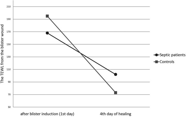
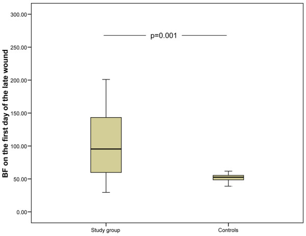
Methods: Blister wounds were induced in 35 patients with severe sepsis and in 15 healthy controls. The healing of the wounds was followed up by measuring transepidermal water loss and blood flow in the wound, reflecting the restoration of the epidermal barrier function and inflammation, respectively. The first set of suction blisters (early wound) was made within 48 hours of the first sepsis-induced organ failure and the second set (late wound) four days after the first wound. In addition, measurements were made on the intact skin.
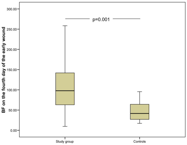
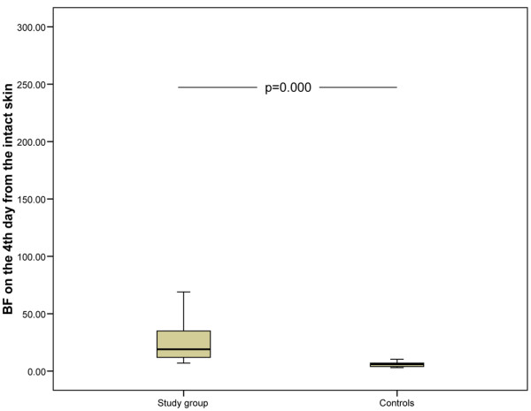
Results: The average age of the whole study population was 62 years (standard deviation [SD] 12). The mean Acute Physiology and Chronic Health Evaluation II (APACHE II) score on admission was 25 (SD 8). The two most common causes of infections were peritonitis and pneumonia. Sixty-six percent of the patients developed multiple organ failure. The decrease in water evaporation from the wound during the first four days was lower in septic patients than in the control subjects (56 g/m2 per hour versus 124 g/m2 per hour, P = 0.004). On the fourth day, septic patients had significantly higher blood flow in the wound compared with the control subjects (septic patients 110 units versus control subjects 47 units, P = 0.001). No difference in transepidermal water loss from the intact skin was found between septic patients and controls. Septic patients had higher blood flow in the intact skin on the fourth and on the eighth day of study compared with the controls.
Conclusions: The restoration of the epidermal barrier function is delayed and wound blood flow is increased in patients with severe sepsis.
Figures
References
-
- Broughton G, 2nd, Janis JE, Attinger CE. Wound healing: an overview. Plast Reconstr Surg. 2006;117:1e-S–32e-S. - PubMed
-
- Laurila JJ, Karttunen T, Koivukangas V, Laurila PA, Syrjälä H, Saarnio J, Soini Y, Ala-Kokko TI. Tight junction proteins in gallbladder epithelium: different expression in acute acalculous and calculous cholecystitis. J Histochem Cytochem. 2007;55:567–573. doi: 10.1369/jhc.6A7155.2007. - DOI - PubMed
MeSH terms
LinkOut - more resources
Full Text Sources
Medical
