. 2009 Apr;39(4):303-14.
doi: 10.1016/j.ibmb.2009.01.007. Epub 2009 Feb 7.

The role of NF-kappaB factor REL2 in the Aedes aegypti immune response

Affiliations

The role of NF-kappaB factor REL2 in the Aedes aegypti immune response

Yevgeniya Antonova et al. Insect Biochem Mol Biol. 2009 Apr.

Abstract

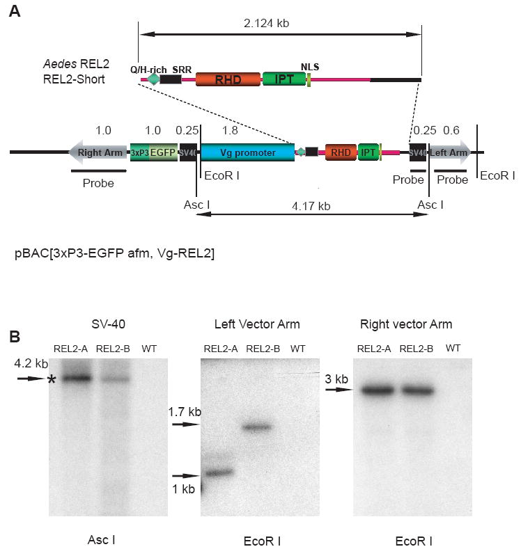
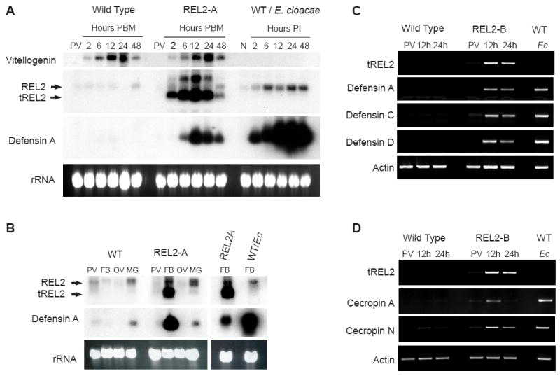
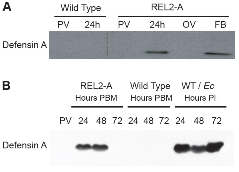
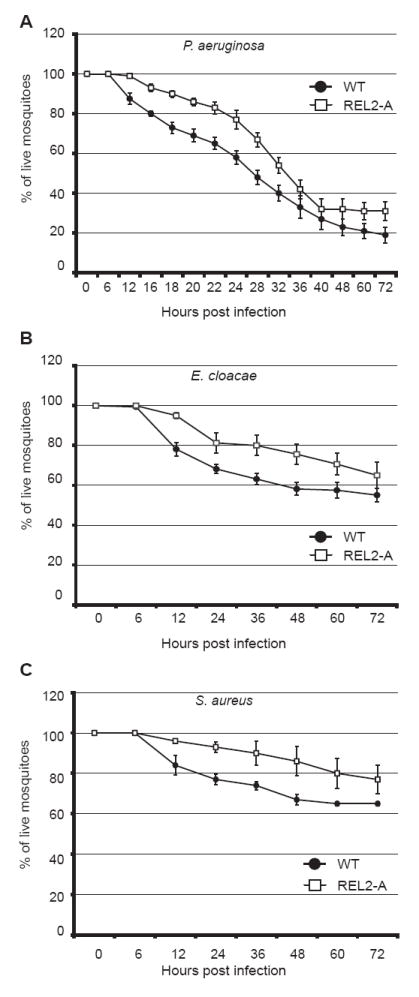
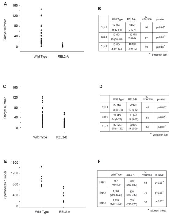
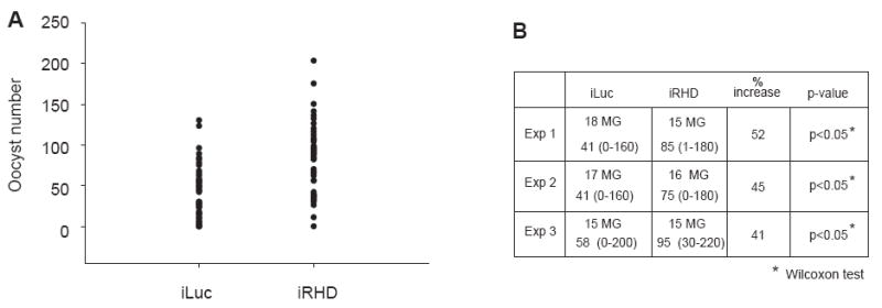
Mosquitoes transmit numerous diseases that continue to be an enormous burden on public health worldwide. Transgenic mosquitoes impervious to vector-borne pathogens, in concert with vector control and drug and vaccine development, comprise an arsenal of means anticipated to defeat mosquito-spread diseases in the future. Mosquito transgenesis allows tissue-specific manipulation of their major immune pathways and enhances the ability to study mosquito-pathogen interactions. Here, we report the generation of two independent transgenic strains of Aedes aegypti overexpressing the NF-?B transcriptional factor REL2, a homologue of Drosophila Relish, which is shown to be under the control of the vitellogenin promoter in the mosquito fat body after a blood meal. We show that this REL2 overexpression in the fat body results in transcriptional activation of Defensins A, C, and D, and Cecropins A and N, as well as translation and secretion of Defensin A protein into the hemolymph. We also demonstrate that induction of REL2 results in the increased resistance of the mosquito to tested Gram-negative and Gram-positive bacteria. Importantly, induction of transgenic REL2 leads to the significant decrease in susceptibility of A. aegypti to Plasmodium gallinaceum infection. Consistently, RNAi knockdown of REL2 in wild-type mosquitoes results in a delay in Defensin A and Cecropin A expression in response to infection and in increased susceptibility to both bacteria and P. gallinaceum. Moreover, our transgenic assays demonstrate that the N-terminus of the mosquito REL2, which includes the His/Gln-rich and serine-rich regions, plays a role in its transactivation properties.


Figures

Figure 1
Figure 1
Generation of two independent transgenic strains of Aedes aegypti—REL2-A and REL2-B (A) Schematic representation of the pBac[3×P3, EGFP afm,Vg-REL2] transformation plasmid used in germ-line transformation. The restriction sites of the restrictases used for DNA digestion are indicated; positions of the probes for right and left vector arms as well as SV40 polyadenylation region are indicated by thick black lines; double-headed arrows indicate the sizes of the REL2-Short isoform (2.124 kb) and the pBAC[3×P3-EGFP afm, Vg-REL2] construct; Q/H, glutamine/histidine-rich region, SRR, serine-rich region, RHD and IPT are parts of the REL DNA-binding domain; NLS, nuclear localization signal. (B) Southern blot analysis of the genomic DNA extracted from REL2-A and REL2-B transgenic mosquito strains and the wild-type strain. Genomic DNA was isolated, digested using EcoR I and ASC I, and hybridized with probes specific to the internal SV40 polyadenylation region, the left and right vector arms; WT—the wild-type parental strain; asterisk indicates the band of expected size.
Figure 2
Figure 2
Blood-meal-activated expression of transgenic REL2 and Defensin and Cecropin families of AMP in the transgenic Ae. aegypti mosquito strains, REL2-A and REL2-B. (A) Northern blot analysis: expression profiles of Vitellogenin, endogenous REL2, transgenic REL2, and Defensin A in the fat body of the REL2-A transgenic mosquitoes. Fat bodies were collected from wild-type (WT) and REL2-A mosquitoes: previtellogenic (PV) and blood-fed mosquitoes at 2, 6, 12, 24, and 48 h post blood meal (PBM). WT mosquitoes were injected with E. cloacae, and the fat bodies were collected from non-infected mosquitoes (n) and infected mosquitoes at 2, 6, 12, 24, and 48 h post-infection (PI). Total RNA was extracted and Northern blot analysis performed using the probes for Vg, REL2 RHD domain, and Defensin A; arrows indicate bands for endogenous REL2—REL2 and transgenic REL2—tREL2, (rRNA) ribosomal RNA—loading control. (B) Northern blot analysis: fat-body-specific expression of transgenic REL2 and Defensin A in REL2-A mosquitoes. Total bodies were collected from previtellogenic (PV) and blood-fed wild-type (WT) and REL2-A mosquitoes at 24 h post blood meal (PBM). Mosquito fat bodies (FB), ovaries (OV), and midguts (MG) were collected from blood-fed mosquitoes at 24 h PBM. Total RNA was extracted, and Northern analysis was performed with the probes for REL2 RHD domain and Defensin A; FB from WT mosquitoes injected with E. cloacae (Ec) at 24 h post-infection were used as a control of Defensin A expression; REL2—endogenous REL2, tREL2—transgenic REL2. (C) Blood-meal-activated expression of transgenic REL2 and Defensins A, C, and D; and (D) blood-meal-activated expression of transgenic REL2 and Cecropin A and N in the transgenic REL2-B strain (RT-PCR analysis). Fat bodies were collected from previtellogenic (PV) and blood-fed mosquitoes at 12 and 24 h post blood meal; fat bodies collected from wild-type (WT) mosquitoes injected with E. cloacae (Ec) at 24 h post-infection were used as a control. Total RNA was extracted and treated with DNase I, and cDNA was synthesized and used as a template for RT-PCR with primers specific for transgenic REL2, Defensin A, C, D, and also Cecropin A and N.
Figure 3
Figure 3
Blood-meal-activated Defensin A protein in the fat bodies (FB) and hemolymph of REL2 transgenic mosquitoes. (A) Blood-meal-activated Defensin A in the FB. Total bodies of previtellogenic (PV) and blood-fed mosquitoes at 24 h post blood meal (PBM), as well as FB and ovaries (OV) of blood-fed REL2-A were used for protein extraction. A 10-μg aliquot of total protein was used per lane for SDS-PAGE, and Western blot analysis was performed using the anti-Defensin A antibodies. (B) Blood-meal-activated secretion of Defensin A peptide in the hemolymph of transgenic mosquitoes. Wild-type and REL2-A mosquitoes were blood fed, and control PV wild-type mosquitoes were injected with E. cloacae; hemolymph was collected as described in Materials and Methods at 24, 48, and 72 h PBM and at the same time points post-infection (PI). Protein (10 μg) was loaded per lane for SDS-PAGE, and Western analysis was performed using the antibodies against Defensin A. WT/Ec—wild-type mosquitoes injected with E. cloacae.
Figure 4
Figure 4
RNAi knockdowns of REL2-Long and REL2-Short result in the delayed expression of AMPs Defensin A and Cecropin A in wild-type, E. cloacae-infected mosquitoes. (A) Expression of REL2-Long and REL2-Short in the REL2 dsRNA-treated mosquitoes. (B and C) Delayed expression of Defensin A (B) and Cecropin A (C) in response to the bacterial challenge in RNAi-treated mosquitoes. iRHD—mosquitoes treated with dsRNA against REL2 RHD domain; iLuc—mosquitoes treated with dsRNA against luciferase gene. N—naïve, non-treated wild-type mosquitoes (neither RNAi nor bacteria treatments); Ecl and E. coli—wild-type mosquitoes not treated with dsRNA, at 24 h post-bacterial infection: EclE. cloacae injections E. coliE. coli injections. Actin amplification represents the loading control for both iRHD and iLuc mosquitoes, where equal amplification of actin was observed in all experiments.
Figure 5
Figure 5
Increase in survival of REL2 transgenics to Gram-negative and Gram-positive bacteria Wild-type and transgenic mosquitoes were blood fed and injected with bacteria between 6 and 12 h post blood meal. Survival was monitored by counting the number of live mosquitoes. (A) Decreased susceptibility of REL2-A transgenics to E. cloacae infection. Combined results of five independent experiments; total number of mosquitoes used was 155; error bars represent standard errors; P values < 0.05 (Cox analysis) (B) Decreased susceptibility of REL2-A transgenics to P. aeruginosa infection. Combined results of six independent experiments; total number of mosquitoes used was 192; error bars represent standard errors; P values < 0.05 (Cox analysis) (C) Decreased susceptibility of REL2-A transgenics to S. aureus infection. Combined results of three independent experiments; total number of mosquitoes used was 96; error bars represent standard errors; P values < 0.05 (Cox analysis)
Figure 6
Figure 6
REL2 RNAi-mediated knockdown results in the increased susceptibility to Gram-negative and Gram-positive bacteria REL2 sdRNA-treated mosquitoes were injected with bacteria on the 4th day after RNAi treatment. Survival was monitored by counting the number of live mosquitoes. (A) Increased susceptibility of iRHD-treated mosquitoes to E. cloacae infection. Combined results of two independent experiments. (B) Increased susceptibility of iRHD mosquitoes to E. faecalis infection; combined results of two independent experiments. iRHD—mosquitoes treated with dsRNA against REL2 RHD domain; iLuc—mosquitoes treated with non-specific dsRNA against luciferase gene; LB—mosquitoes injected with sterile LB media.
Figure 7
Figure 7
Increased resistance of REL2 transgenic Ae. aegypti to P. gallinaceum infection. (A) and (C) Decreased number of P. gallinaceum oocysts in the midguts of REL2-A and REL2-B mosquitoes, respectively. (B) and (D) Numeric representation of the graphs in (A) and (C), respectively; average oocyst number for REL2-A, REL2-B, and wild-type mosquitoes represents the average range of number of oocysts per experiment (shown in parentheses); number of midguts used per experiment is indicated; (MG) midguts. (E) and (F) Sporozoite number was significantly reduced in REL2-A mosquitoes: (C) P. gallinaceum sporozoite number in the REL2-A mosquitoes; (D) numeric representation of the graph in (C); the table reflects average number of sporozoites and the range is indicated in parentheses.
Figure 8
Figure 8
REL2 RNAi-mediated knockdown results in the increased susceptibility of Ae. aegypti to P. gallinaceum infection. (A) Increased number of P. gallinaceum oocysts in the midguts of iRHD-treated mosquitoes versus control iLuc mosquitoes (P < 0.05 Wilcoxon test). (B) Numeric representation of the graph in (A), average oocyst number for REL2 RNAi (iREL2), and control Luciferase RNAi (iLuc) mosquitoes represents the average range of number of oocysts per experiment (shown in parentheses). Number of midguts used per experiment is indicated; (MG) midguts.

Similar articles

Cited by

References

    1. Apidianakis Y, Mindrinos MN, Xiao W, Lau GW, Baldini RL. Profiling early infection responses: Pseudomonas aeruginosa eludes host defenses by suppressing antimicrobial peptide gene expression. Proc Natl Acad Sci U S A. 2005;102:2573–2578. - PMC - PubMed
    1. Baeuerle PA, Baltimore D. NF-κB: Ten Years After. Cell. 1996;87:13–20. - PubMed
    1. Bian G, Shin SW, Cheon HM, Kokoza V, Raikhel AS. Transgenic alteration of Toll immune pathway in the female mosquito Aedes aegypti. Proc Natl Acad Sci U S A. 2005;102:13568–13573. - PMC - PubMed
    1. Blandin S, Moita KF, Kocher T, Wilm M, Kafatos FC, Levashina EA. Reverse genetics in the mosquito Anopheles gambiae: targeted disruption of the defensin gene. EMBO Rep. 2002;3:852–856. - PMC - PubMed
    1. Christophides GK, Zdobnov E, Barillas-Mury C, Birney E, Blandin S, Blass C, Brey PT, Collins FH, Danielli A, Dimopoulos G, Hetru C, Hoa NT, Hoffmann JA, Kanzok SM, Letunic I, Levashina EA, Loukeris TG, Lycett G, Meister S, Michel K, Moita LF, Müller HM, Osta MA, Paskewitz SM, Reichhart JM, Rzhetsky A, Troxler L, Vernick KD, Vlachou D, Volz J, von Mering C, Xu J, Zheng L, Bork P, Kafatos FC. Immunity-related genes and gene families in Anopheles gambiae. Science. 2002;298:59–65. - PubMed

Publication types

MeSH terms

LinkOut - more resources