. 2009 Jul;63(6):429-35.
doi: 10.1016/j.biopha.2009.04.045. Epub 2009 Jun 9.

JNK/AP-1 pathway is involved in tumor necrosis factor-alpha induced expression of vascular endothelial growth factor in MCF7 cells

Affiliations

JNK/AP-1 pathway is involved in tumor necrosis factor-alpha induced expression of vascular endothelial growth factor in MCF7 cells

Yongmei Yin et al. Biomed Pharmacother. 2009 Jul.

Abstract

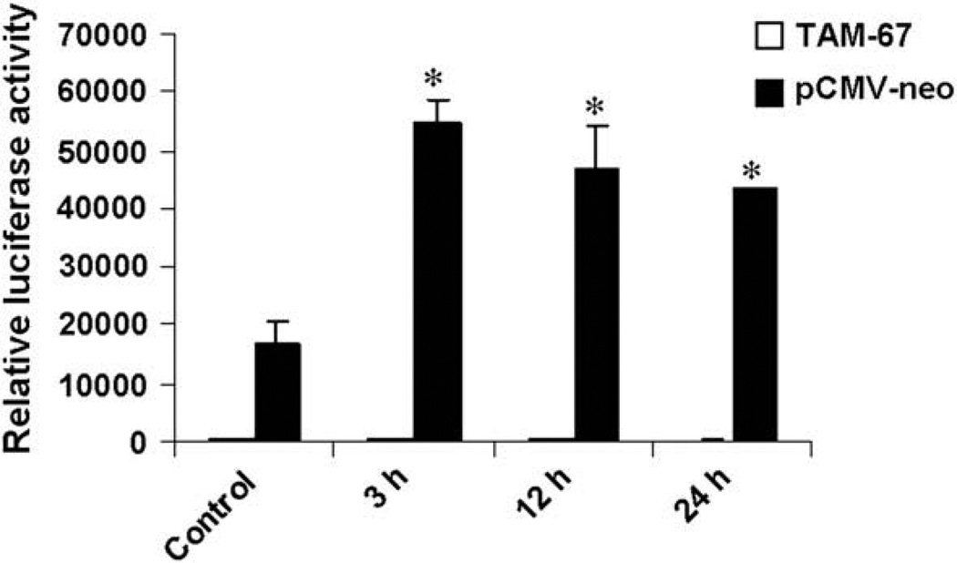
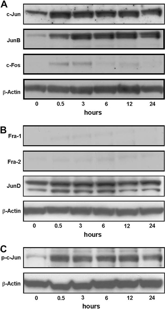
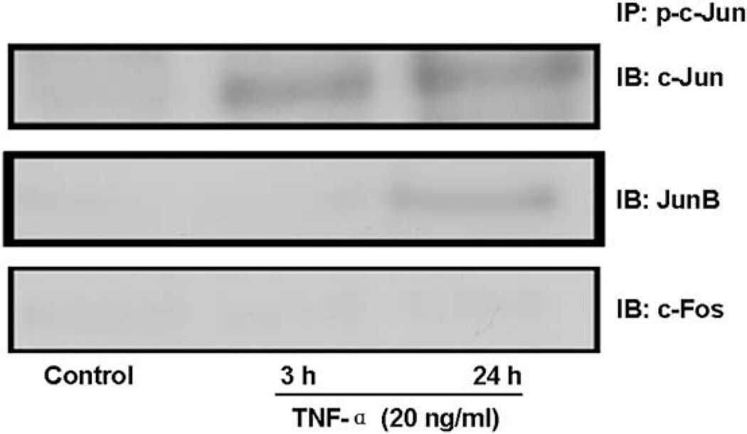
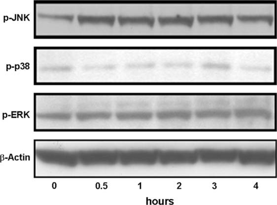
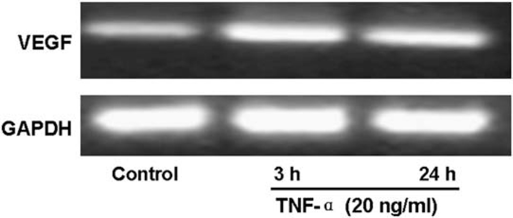
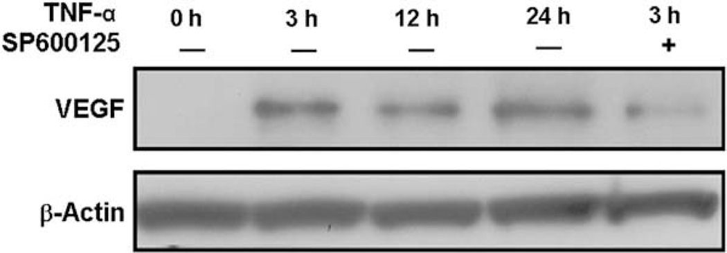
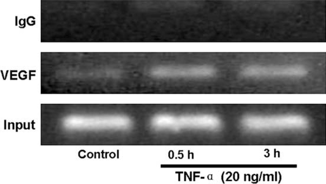
Vascular endothelial growth factor (VEGF) has been implicated in breast tumor angiogenesis. And tumor necrosis factor-alpha (TNF-alpha) is a positive regulator of VEGF. This study was aimed to identify the signalling pathway of TNF-alpha in VEGF expression regulation in breast cancer cell line MCF7. Using luciferase reporter assays, we demonstrated that TNF-alpha significantly increased activator protein-1 (AP-1) transcriptional activity in the MCF7 cells. The expression of the AP-1 family members c-Jun, c-Fos and JunB and phosphorylation levels of c-Jun were upregulated by TNF-alpha, whereas other AP-1 family members Fra-1, Fra-2, and JunD were unaffected. The activation of AP-1 was associated with the formation of p-c-Jun-c-Jun and p-c-Jun-JunB homodimers. Furthermore, the phosphorylation levels of c-Jun N-terminal kinase (JNK) but not P38 and ERK were elevated by TNF-alpha in MCF7 cells. TNF-alpha potently upregulated the mRNA and protein levels of VEGF, which were significantly reversed by JNK inhibitor SP600125. Finally using chromatin immunoprecipitation (CHIP) assays, we found that p-c-Jun bound to the VEGF promoter and regulated VEGF transcription directly. These data suggest that the pro-inflammatory cytokine TNF-alpha is a critical regulator of VEGF expression in breast cancer cells, at least partially via a JNK and AP-1 dependent pathway.


Figures

Fig. 1
Fig. 1
TNF-α elevates AP-1 luciferase reporter activity. MCF7 cells were transiently cotransfected with the firefly luciferase reporter construct AP-1-LUC and β-galactosidase plasmid (as an internal control) plus pCMV-TAM-67 or pCMV-neo vector. Eight hours after transfection, cells were serum starved for 24 h, and then treated with TNF-α (20 ng/ml) or vehicle (Control) for the indicated periods of time. Then cells were harvested for luciferase reporter assays. The relative values of AP-1 luciferase activity to β-galactosidase are shown as means ± SEM of three independent experiments. *P < 0.05 vs. levels in cells cotransfected with pCMV-neo vector and treated with vehicle.
Fig. 2
Fig. 2
TNF-α induces the expression of c-Jun, JunB and c-Fos, but not other Jun or Fos proteins. MCF7 cells were serum starved for 24 h and treated with TNF-α (20 ng/ml) for indicated periods of time. Nuclear protein samples were harvested for Western blot analyses to determine the protein levels of c-Jun, JunB, c-Fos (A), Fra-1, Fra-2, JunD (B) and Ser73-phosphorylated-c-Jun (p-c-Jun; C). The protein levels of β-actin were assessed simultaneously in each group as a loading control. Immunoblots are representative of three separate experiments.
Fig. 3
Fig. 3
AP-1 complex is composed of p-c-Jun-c-Jun and p-c-Jun-JunB homodimers after TNF-α treatment. MCF7 cells were serum starved for 24 h and treated without (Control) or with TNF-α (20 ng/ml) for 3 h or 24 h. Cell nuclear lysates were subjected to immunoprecipitation (IP) with an antibody against p-c-Jun followed by immunoblotting (IB) with antibodies against c-Jun, c-Fos and JunB. Representative Immunoblots of three separate experiments are shown.
Fig. 4
Fig. 4
Effects of TNF-α on the activities of MAPK pathways. MCF7 cells were serum starved for 24 h and treated with TNF-α (20 ng/ml) for the indicated periods of time, and JNK (p-JNK), P38 (p-p38) and ERK (p-ERK) activation was assessed using phosphorylation specific antibodies. The levels of β-actin protein in each group were used as the references. Representative immunoblots of three separate experiments are shown.
Fig. 5
Fig. 5
TNF-α induces VEGF luciferase reporter activity. MCF7 cells were transiently transfected with VEGF-LUC construct and β-galactosidase plasmid (as an internal control). Eight hours after transfection, cells were serum starved for 24 h, and then treated with TNF-α (20 ng/ml) or vehicle (Control) for the indicated periods of time. Then cells were collected for luciferase reporter assays. The relative values of LXRE luciferase activity to β-galactosidase are shown as means ± SEM of three independent experiments. *P < 0.05 vs. control.
Fig. 6
Fig. 6
TNF-α stimulates VEGF mRNA transcription in MCF7 cells. MCF7 cells were treated with 20 ng/ml of TNF-α for the indicated time periods after 24 h serum starvation. Total RNA was isolated and 1 µg RNA was used for RT-PCR analyses of VEGF and GAPDH (as a reference) mRNA levels. PCR products were revealed by ethidium bromide staining under UV after agarose gel electrophoresis. Representative graphs of three separate experiments are shown.
Fig. 7
Fig. 7
TNF-α increases VEGF protein levels in MCF7 cells. After serum starvation for 24 h, MCF7 cells were pretreated without or with SP600125 for 30 min, and treated with TNF-α (20 ng/ml) for the indicated time periods. Cell lysates were collected for Western blot analyses with antibodies against VEGF or β-actin (as a loading control). Representative Immunoblots of three separate experiments are shown.
Fig. 8
Fig. 8
p-c-Jun is recruited to the VEGF promoter following TNF-α treatment. After serum starvation for 24 h, MCF7 cells were treated without (Control) or with TNF-α (20 ng/ml) for the indicated time periods, and chromatin protein-DNA complexes were cross-linked using formaldehyde. The purified nucleoprotein complexes were immunoprecipitated with Ser73-phosphorylated-c-Jun antibody (p-c-Jun) or nonimmune IgG and DNA samples recovered were amplified by PCR as detailed in Materials and Methods. Representative graphs of three separate experiments are shown.

References

    1. Ferrara N, Davis-Smyth T. The biology of vascular endothelial growth factor. Endocr Rev. 1997;18(1):4–25. - PubMed
    1. Leek RD, Harris AL, Lewis CE. Cytokine networks in solid human tumors: regulation of angiogenesis. J Leukoc Biol. 1994;56(4):423–435. - PubMed
    1. Clauss M, Gerlach M, Gerlach H, Brett J, Wang F, Familletti PC, et al. Vascular permeability factor: a tumor-derived polypeptide that induces endothelial cell and monocyte procoagulant activity, and promotes monocyte migration. J Exp Med. 1990;172(6):1535–1545. - PMC - PubMed
    1. Tischer E, Mitchell R, Hartman T, Silva M, Gospodarowicz D, Fiddes JC, et al. The human gene for vascular endothelial growth factor. Multiple protein forms are encoded through alternative exon splicing. J Biol Chem. 1991;266(18):11947–11954. - PubMed
    1. Forsythe JA, Jiang BH, Iyer NV, Agani F, Leung SW, Koos RD, et al. Activation of vascular endothelial growth factor gene transcription by hypoxia-inducible factor 1. Mol Cell Biol. 1996;16(9):4604–4613. - PMC - PubMed

Publication types

MeSH terms