

Linking molecular affinity and cellular specificity in cadherin-mediated adhesion
- PMID: 19553217
- PMCID: PMC2710653
- DOI: 10.1073/pnas.0905349106
Linking molecular affinity and cellular specificity in cadherin-mediated adhesion
Abstract
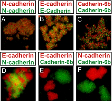
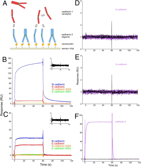
Many cell-cell adhesive events are mediated by the dimerization of cadherin proteins presented on apposing cell surfaces. Cadherin-mediated processes play a central role in the sorting of cells into separate tissues in vivo, but in vitro assays aimed at mimicking this behavior have yielded inconclusive results. In some cases, cells that express different cadherins exhibit homotypic cell sorting, forming separate cell aggregates, whereas in other cases, intermixed aggregates are formed. A third pattern is observed for mixtures of cells expressing either N- or E-cadherin, which form distinct homotypic aggregates that adhere to one another through a heterotypic interface. The molecular basis of cadherin-mediated cell patterning phenomena is poorly understood, in part because the relationship between cellular adhesive specificity and intermolecular binding free energies has not been established. To clarify this issue, we have measured the dimerization affinities of N-cadherin and E-cadherin. These proteins are similar in sequence and structure, yet are able to mediate homotypic cell patterning behavior in a variety of tissues. N-cadherin is found to form homodimers with higher affinity than does E-cadherin and, unexpectedly, the N/E-cadherin heterophilic binding affinity is intermediate in strength between the 2 homophilic affinities. We can account for observed cell aggregation behaviors by using a theoretical framework that establishes a connection between molecular affinities and cell-cell adhesive specificity. Our results illustrate how graded differences between different homophilic and heterophilic cadherin dimerizaton affinities can result in homotypic cell patterning and, more generally, show how proteins that are closely related can, nevertheless, be responsible for highly specific cellular adhesive behavior.
Conflict of interest statement
The authors declare no conflict of interest.
Figures
References
-
- Takeichi M. Cadherins: A molecular family important in selective cell-cell adhesion. Annu Rev Biochem. 1990;59:237–252. - PubMed
-
- Halbleib JM, Nelson WJ. Cadherins in development: cell adhesion, sorting, and tissue morphogenesis. Genes Dev. 2006;20:3199–3214. - PubMed
-
- Duguay D, Foty RA, Steinberg MS. Cadherin-mediated cell adhesion and tissue segregation: Qualitative and quantitative determinants. Dev Biol. 2003;253:309–323. - PubMed
-
- Nollet F, Kools P, van Roy F. Phylogenetic analysis of the cadherin superfamily allows identification of six major subfamilies besides several solitary members. J Mol Biol. 2000;299:551–572. - PubMed
Publication types
MeSH terms
Substances
Grants and funding
LinkOut - more resources
Full Text Sources
Molecular Biology Databases
Research Materials
