. 2009 Sep;83(17):8655-61.
doi: 10.1128/JVI.01129-09. Epub 2009 Jun 24.

Palmitoylation of the influenza A virus M2 protein is not required for virus replication in vitro but contributes to virus virulence

Affiliations

Palmitoylation of the influenza A virus M2 protein is not required for virus replication in vitro but contributes to virus virulence

Michael L Grantham et al. J Virol. 2009 Sep.

Abstract

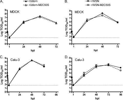
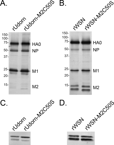
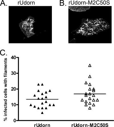
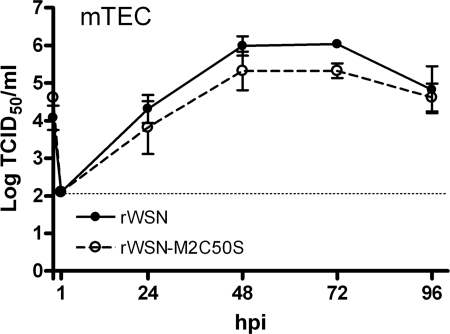
The influenza A virus M2 protein has important roles during virus entry and in the assembly of infectious virus particles. The cytoplasmic tail of the protein can be palmitoylated at a cysteine residue, but this residue is not conserved in a number of human influenza A virus isolates. Recombinant viruses encoding M2 proteins with a serine substituted for the cysteine at position 50 were generated in the A/WSN/33 (H1N1) and A/Udorn/72 (H3N2) genetic backgrounds. The recombinant viruses were not attenuated for replication in MDCK cells, Calu-3 cells, or in primary differentiated murine trachea epithelial cell cultures, indicating there was no significant contribution of M2 palmitoylation to virus replication in vitro. The A/WSN/33 M2C50S virus displayed a slightly reduced virulence after infection of mice, suggesting that there may be novel functions for M2 palmitoylation during in vivo infection.


Figures

FIG. 1.
FIG. 1.
Altering the palmitoylation site of M2 does not affect infectious virus production in tissue culture cells. MDCK cells (A and B) or polarized Calu-3 cells (C and D) were infected with the indicated viruses. The amount of infectious virus in cell supernatants harvested at the indicated times was determined by TCID50 assay. Data points indicate the average and error bars represent the standard error of the mean from triplicate samples. The horizontal dotted lines represent the limits of detection.
FIG. 2.
FIG. 2.
Altering the palmitoylation site of M2 does not affect the protein composition of virions. Virus particles concentrated by ultracentrifugation through a 20% sucrose cushion were analyzed by Western blotting for HA, NP, M1 (A and B), and M2 incorporation (C and D). Molecular weight standards are shown to the left of each blot (A and B).
FIG. 3.
FIG. 3.
Alteration of the M2 palmitoylation site does not affect filamentous virus particle formation. The ability of rUdorn or rUdorn-M2C50S to form filaments was examined by immunofluorescence assay for HA followed by confocal microscopy. Panels A and B show projections of z stacks taken at ×63 magnification. The percentage of infected cells with filaments was also determined for a total of 20 fields viewed with a ×20 objective (C).
FIG. 4.
FIG. 4.
Altering the palmitoylation site of M2 attenuates virus virulence in vivo. Mice were infected intranasally with the indicated doses of rWSN or rWSN-M2C50S and monitored for 14 days for mortality (A) and weight loss (B and C). Each group contained 10 animals except for those infected with 103 TCID50, which contained 5 animals. Body mass p.i. was normalized to mass at the time of infection. Error bars indicate the standard error of the mean. The lungs from infected mice were homogenized, and the amount of infectious virus (D), MIP-1α (E), and IL-10 (F) per gram of tissue was determined. An asterisk indicates a P value of <0.05. The horizontal lines in the graphs represent mean titers.
FIG. 5.
FIG. 5.
Alteration of the M2 palmitoylation site does not affect virus replication in primary, differentiated mTEC. mTEC cultures were infected with the indicated viruses, and at the indicated times the apical supernatants were harvested. The amount of infectious virus in each sample was determined by TCID50 assay on MDCK cells. Data points indicate the average from two independent experiments each done in duplicate. Error bars indicate the standard error of the mean, and the dotted line indicates the limit of detection.

Similar articles

Cited by

References

    1. Benton, K. A., J. A. Misplon, C. Y. Lo, R. R. Brutkiewicz, S. A. Prasad, and S. L. Epstein. 2001. Heterosubtypic immunity to influenza A virus in mice lacking IgA, all Ig, NKT cells, or gamma delta T cells. J. Immunol. 1667437-7445. - PubMed
    1. Castrucci, M. R., M. Hughes, L. Calzoletti, I. Donatelli, K. Wells, A. Takada, and Y. Kawaoka. 1997. The cysteine residues of the M2 protein are not required for influenza A virus replication. Virology 238128-134. - PubMed
    1. Centers for Disease Control and Prevention. 2009. Swine influenza A (H1N1) infection in two children—Southern California, March-April 2009. MMWR Morb. Mortal. Wkly. Rep. 58400-402. - PubMed
    1. Chen, B. J., G. P. Leser, D. Jackson, and R. A. Lamb. 2008. The influenza virus M2 protein cytoplasmic tail interacts with the M1 protein and influences virus assembly at the site of virus budding. J. Virol. 8210059-10070. - PMC - PubMed
    1. Chen, B. J., M. Takeda, and R. A. Lamb. 2005. Influenza virus hemagglutinin (H3 subtype) requires palmitoylation of its cytoplasmic tail for assembly: M1 proteins of two subtypes differ in their ability to support assembly. J. Virol. 7913673-13684. - PMC - PubMed

Publication types

MeSH terms

LinkOut - more resources