. 2009 Sep;83(17):8744-58.
doi: 10.1128/JVI.00613-09. Epub 2009 Jun 24.

Proteolytic activation of the spike protein at a novel RRRR/S motif is implicated in furin-dependent entry, syncytium formation, and infectivity of coronavirus infectious bronchitis virus in cultured cells

Affiliations

Proteolytic activation of the spike protein at a novel RRRR/S motif is implicated in furin-dependent entry, syncytium formation, and infectivity of coronavirus infectious bronchitis virus in cultured cells

Yoshiyuki Yamada et al. J Virol. 2009 Sep.

Abstract

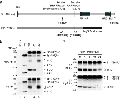
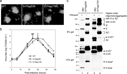
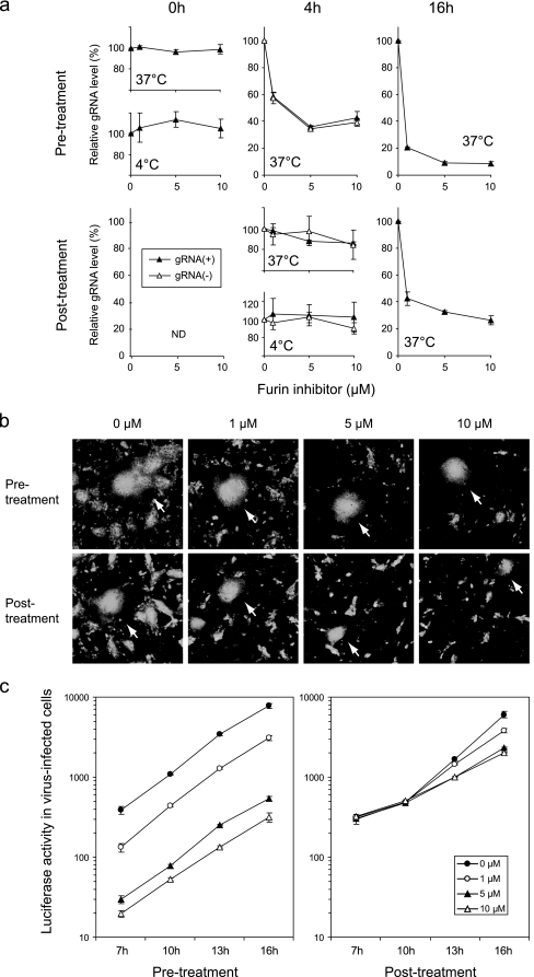
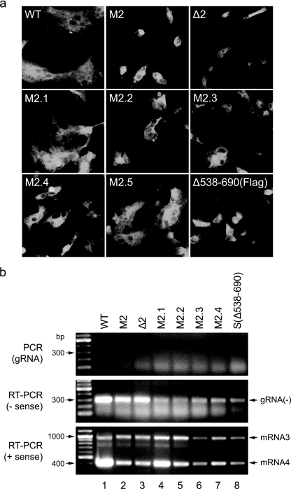
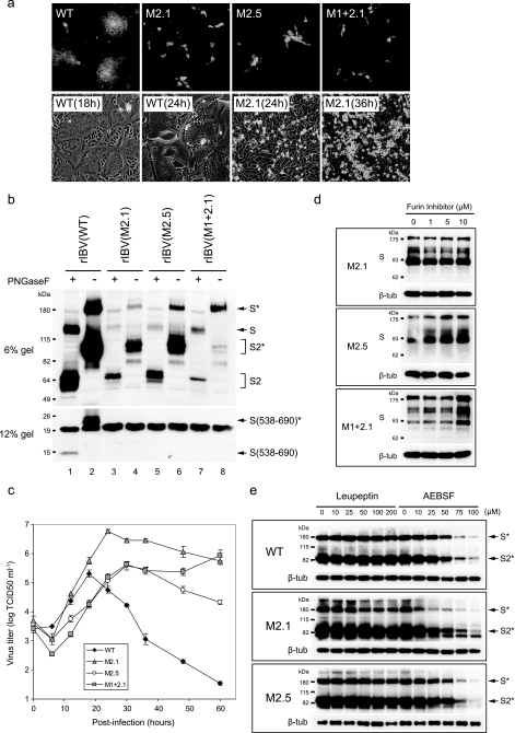
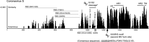
The spike (S) protein of the coronavirus (CoV) infectious bronchitis virus (IBV) is cleaved into S1 and S2 subunits at the furin consensus motif RRFRR(537)/S in virus-infected cells. In this study, we observe that the S2 subunit of the IBV Beaudette strain is additionally cleaved at the second furin site (RRRR(690)/S) in cells expressing S constructs and in virus-infected cells. Detailed time course experiments showed that a peptide furin inhibitor, decanoyl-Arg-Val-Lys-Arg-chloromethylketone, blocked both viral entry and syncytium formation. Site-directed mutagenesis studies revealed that the S1/S2 cleavage by furin was not necessary for, but could promote, syncytium formation by and infectivity of IBV in Vero cells. In contrast, the second site is involved in the furin dependence of viral entry and syncytium formation. Mutations of the second site from furin-cleavable RRRR/S to non-furin-cleavable PRRRS and AAARS, respectively, abrogated the furin dependence of IBV entry. Instead, a yet-to-be-identified serine protease(s) was involved, as revealed by protease inhibitor studies. Furthermore, sequence analysis of CoV S proteins by multiple alignments showed conservation of an XXXR/S motif, cleavable by either furin or other trypsin-like proteases, at a position equivalent to the second IBV furin site. Taken together, these results suggest that proteolysis at a novel XXXR/S motif in the S2 subunit might be a common mechanism for the entry of CoV into cells.


Figures

FIG. 1.
FIG. 1.
An in vitro assay shows that RRRR690/S of the IBV S protein is a furin substrate. (a) Schematic diagram of S protein from Vero-adapted IBV Beaudette and furin cleavage sites predicted by the ProP server. Constructs that encode full-length S and truncated S fused to the human IgG Fc domain, S(1-789)Fc, are illustrated. The positions for inserting the Flag tag, the putative signal sequence (SS), the fusion peptide (FP), heptad repeats 1 and 2 (HR1 and HR2), the transmembrane domain (TM), and the amino acid (aa) sequences deleted in the two deletion constructs (Δ1 and Δ2) are also shown. (b) Detection of cleavage at the two furin sites. The S(1-789)Fc constructs were expressed in HuH-7 cells for 24 h, total cell lysates were prepared, and PNGase F (New England Biotechnology) treatment was carried out for 2 h at 37°C. Cell extracts were analyzed by WB. (c) Inhibition of furin-dependent cleavage by furin inhibitor. At 6 h posttransfection of S(1-789)Fc constructs in HuH-7 cells, the culture media were replaced with fresh DMEM containing different concentrations of furin inhibitor. The cells were further incubated for 18 h, and cell extracts were analyzed by WB.
FIG. 2.
FIG. 2.
Cleavage products at two furin sites are detected in IBV-infected cells. (a) Syncytium formation by rIBV was not affected by Flag-tagged S genes. Vero cells were infected with rIBV for 2 h. The cells were washed three times with PBS and incubated for 16 h with an overlay of DMEM containing 0.6% carboxymethyl cellulose. Syncytium formation was observed by IF staining with the S2 Ab. (b) Replication of S-Flag1163 was slightly delayed compared to the WT and S-Flag539. Vero cells (in a 25-cm2 flask) were infected with rIBV and harvested at 0, 6, 12, 18, 24, 30, 36, 42, and 48 h postinfection. Viral stocks were prepared by freezing/thawing of the cells three times, and the TCID50 was determined. The arrows indicate the time points when CPE was observed in nearly 100% of the cells. The error bars indicate standard errors of the means. (c) Cleavage at two furin sites was detected in virus-infected cells. HuH-7 cells were infected with rIBV for 18 h. Cell extracts were analyzed by WB with the anti-Flag Ab.
FIG. 3.
FIG. 3.
Entry and syncytium formation of IBV are blocked by furin inhibitor. (a) Pretreatment with furin inhibitor did not affect attachment but blocked IBV entry. The gRNA level in virus-infected cells was investigated with a quantitative real-time RT-PCR. The values are shown as relative amounts against 0 μM of furin inhibitor treatment. ND, not done. The error bars indicate standard deviations of the means. (b) The number of infected cells was reduced by pretreatment with furin inhibitor, while posttreatment blocked giant syncytium formation. Syncytium formation was observed by IF staining with the S2 Ab at 16 h postinfection. Giant syncytia caused by primary infection are indicated by arrows. (c) Inhibitory effects of pretreatment with furin inhibitor were caused by less primary infection, while those of posttreatment were due to blocking of cell-to-cell spread and secondary infection. The effects of furin inhibitor treatments on rIBV-Luc infection were investigated. Luciferase activity in virus-infected cells was measured at 7, 10, 13, and 16 h postinfection. The error bars indicate standard deviations of the means.
FIG. 4.
FIG. 4.
S1/S2 cleavage by furin is not essential for, but could promote, syncytium formation and infectivity of IBV. (a) Cleavage deficiency at the S1/S2 site resulted in reduced size of syncytia. Syncytium formation was observed as described in the legend to Fig. 2a. (b) A minute amount of S1/S2 cleavage was detected in M1- and Δ1-infected cells, although they do not have a furin consensus motif. HuH-7 cells were infected with rIBV for 18 h. Cell extracts (20 μg protein) were analyzed by immunoblotting them with the S2 Ab. (c) Viral replication and spread were reduced in the absence of S1/S2 cleavage. Growth kinetics was investigated as described in the legend to Fig. 2b. The arrow indicates the time point when CPE was observed in nearly 100% of the cells. The error bars indicate standard deviations of the means. (d) Infection of M1 and Δ1 were inhibited by pretreatment with furin inhibitor. Vero cells were pretreated with different concentrations of furin inhibitor and infected with rIBV for 2 h. The cells were washed three times with PBS and incubated with fresh DMEM without furin inhibitor. Replication of viruses was observed by immunoblotting them with the S2 Ab at 16 h postinfection. β-tub, β-tubulin.
FIG. 5.
FIG. 5.
Infectivity to Vero cells was abrogated by mutations of the second furin site in S protein. (a) Trypsin activation of cell-cell fusion was inefficient in the second furin site mutants, which cannot recover infectious viruses. Trypsin (2 μg/ml) was added at 10 h posttransfection of S constructs, and the cells were incubated for 2 h at 37°C. IF staining was carried out with either anti-S2 or anti-Flag Ab. (b) Replication and transcription of viral RNAs were observed in cells electroporated with mutant transcripts, although the infectious clones were not recovered. At 48 h postelectroporation of the full-length transcripts, total RNA was extracted from the cells. Negative-strand gRNA and subgenomic mRNA were detected by RT-PCR amplification.
FIG. 6.
FIG. 6.
Proteolytic activation at the second IBV furin site is mediated by furin and other serine protease(s). (a) Giant syncytium formation by rIBV was mediated by proteolytic activation at the second furin site. Syncytium formation was observed as described in the legend to Fig. 2a. For observation of CPE, cells were incubated with DMEM instead of overlay medium. (b) S1/S2 cleavage occurred efficiently in M2.1- and M2.5-infected cells, but not in M1+2.1-infected cells. HuH-7 cells were infected with rIBV for 18 h. Cell extracts (20 μg protein) were analyzed by immunoblotting them with the S2 Ab. (c) M2.1 and M1+2.1, which lack induction of syncytia, demonstrated prolonged replication with high productivity of virions. The growth kinetics was investigated as described in the legend to Fig. 2b. The error bars indicate standard deviations of the means. (d) Infection of M2.1, M1+2.1, and M2.5 was not affected by pretreatment with furin inhibitor. The effect of pretreatment with furin inhibitor was investigated as described in the legend to Fig. 4d. (e) Infection of the WT, M2.1, and M2.5 was inhibited by pretreatment with AEBSF, but not leupeptin. The effects of protease inhibitors were investigated as described in the legend to Fig. 4d, except that the cells were pretreated with leupeptin or AEBSF.
FIG. 7.
FIG. 7.
A novel XXXR/S motif, cleavable by furin, general PCs, or trypsin, is strictly conserved in CoV. The similarity alignments of CoV S were obtained with the Vector NTI Align X software (Informax). The fusion peptide (FP), heptad repeats 1 and 2 (HR1 and HR2), and the transmembrane domain (TM) are also shown. The gaps in the HR1 and HR2 regions were due to amino acid insertion in a group 1 CoV. Receptor-binding domains (RBD) are shown with dashed lines. The S1/S2 cleavage and the second IBV furin site are indicated by arrows. The asterisk indicates the site of S1/S2 cleavage by furin in group 1 (feline CoV Black and NTU2), group 2 (MHV, bovine CoV, human CoV OC43 and HKU-1, porcine hemagglutinating encephalomyelitis virus, and equine CoV), and group 3 (IBV and turkey CoV). S1/S2 cleavage in SARS-CoV by trypsin (empty arrow) and cathepsin (solid arrow) is also shown.

Similar articles

Cited by

References

    1. Alonso-Caplen, F. V., Y. Matsuoka, G. E. Wilcox, and R. W. Compans. 1984. Replication and morphogenesis of avian coronavirus in Vero cells and their inhibition by momensin. Virus Res. 1153-167. - PMC - PubMed
    1. Basak, A., M. Zhong, J. S. Munzer, M. Chretien, and N. G. Seidah. 2001. Implication of the proprotein convertases furin, PC5 and PC7 in the cleavage of surface glycoproteins of Hong Kong, Ebola and respiratory syncytial viruses: a comparative analysis with fluorogenic peptides. Biochem. J. 353537-545. - PMC - PubMed
    1. Bergeron, E., M. J. Vincent, L. Wickham, J. Hamelin, A. Basak, S. T. Nichol, M. Chretien, and N. G. Seidah. 2005. Implication of proprotein convertases in the processing and spread of severe acute respiratory syndrome coronavirus. Biochem. Biophys. Res. Commun. 326554-563. - PMC - PubMed
    1. Bonavia, A., B. D. Zelus, D. E. Wentworth, P. J. Talbot, and K. V. Holmes. 2003. Identification of a receptor-binding domain of the spike glycoprotein of human coronavirus HCoV-229E. J. Virol. 772530-2538. - PMC - PubMed
    1. Bos, E. C., L. Heijnen, W. Luytjes, and W. J. Spaan. 1995. Mutational analysis of the murine coronavirus spike protein: effect on cell-to-cell fusion. Virology 214453-463. - PMC - PubMed

Publication types

LinkOut - more resources