. 2009 Sep;83(17):8849-58.
doi: 10.1128/JVI.00339-09. Epub 2009 Jun 24.

Kaposi's sarcoma-associated herpesvirus protein LANA2 disrupts PML oncogenic domains and inhibits PML-mediated transcriptional repression of the survivin gene

Affiliations

Kaposi's sarcoma-associated herpesvirus protein LANA2 disrupts PML oncogenic domains and inhibits PML-mediated transcriptional repression of the survivin gene

Laura Marcos-Villar et al. J Virol. 2009 Sep.

Abstract

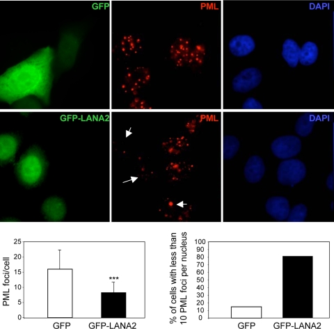
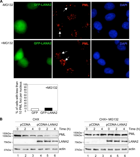
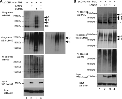
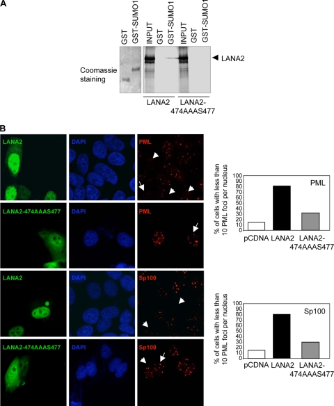
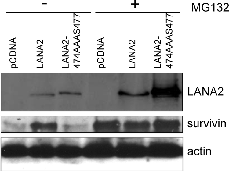
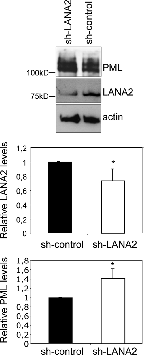
Infection by herpesviruses causes a dramatic disturbance of PML oncogenic domains (PODs) that has been suggested to be essential for viral lytic replication. Several proteins from Kaposi's sarcoma-associated herpesvirus (KSHV) have been tested as putative POD-disrupting factors with negative results. Here, we show that LANA2, a viral protein that is absolutely required for the viability and proliferation of KSHV-infected primary effusion lymphoma (PEL) cells, increases the levels of SUMO2-ubiquitin-modified PML and induces the disruption of PODs by a proteasome-mediated mechanism. In addition, we demonstrate that this disruption is largely dependent on both the integrity of a SUMO interaction motif in LANA2 and the lysine 160 from PML. Moreover, silencing of LANA2 expression in PEL cells by RNA interference led to an increase in the PML levels. Finally, we demonstrate that LANA2 relieves PML-mediated transcriptional repression of survivin, a protein that directly contributes to malignant progression of PEL. This represents the first example of inactivation of these important antiviral structures by KSHV.


Figures

FIG. 1.
FIG. 1.
LANA2 induces the displacement of PML from PODs. MCF-7 cells were transfected as indicated and stained with anti-PML antibody. Arrows indicate cells that are positively expressing LANA2. The number of PML dots per cell as well as the percentage of cells with fewer than 10 PML dots is presented, counting at least 200 transfected cells. ***, P < 0.0005, compared with cells not expressing LANA2 (Student's test).
FIG. 2.
FIG. 2.
Disruption of PODs and degradation of PML by LANA2 are mediated by the proteasome. (A) Disruption of PODs by LANA2 is mediated by the proteasome. MCF-7 cells were transfected and treated with the proteasome inhibitor MG132 as indicated. Endogenous PML was detected by immunofluorescence using anti-PML antibody. The weaker speckled pattern detected in cells expressing GFP-LANA2 is recovered after MG132 treatment. Arrows indicate cells that are positively expressing LANA2. The percentage of transfected cells with fewer than 10 PML dots is presented, counting at least 200 transfected cells. (B) LANA2 expression induces the degradation of HA-PML by a proteasome-dependent mechanism. Cells cotransfected with HA-PMLIV and pCDNA or pCDNA-LANA2 were treated with cycloheximide (CHX) in the presence or absence of the proteasome inhibitor MG312 and harvested at the indicated time points. Cell extracts were then analyzed by Western blotting with the indicated antibodies.
FIG. 3.
FIG. 3.
PML residue lysine 160 is required for the disruption of PODs induced by LANA2. MCF-7 cells were transiently transfected with pCDNA or pCDNA-LANA2 plasmid together with plasmids that encoded PML WT or the indicated PML mutants and stained for LANA2 (green) and PML (red). PML-3KR, PML with arginine residues replacing the three lysine residues that covalently bind to SUMO.
FIG. 4.
FIG. 4.
Upregulation of SUMO2 and ubiquitin modification of PML by LANA2. (A) MCF-7 cells were transfected with the indicated plasmids, and at 60 h after transfection His-tagged PML protein was purified from the cell extracts by nickel affinity chromatography before analysis. Purified extracts were then analyzed by Western blotting (WB) using antibodies to SUMO2, PML, or ubiquitin (Ub). Input extracts were incubated with anti-LANA2 or anti-actin antibodies. A shorter exposition of the gel is shown in the right-hand panel. (B) MCF-7 cells were transfected with the indicated plasmids, and 36 h after transfection His-tagged PML protein was purified from the cell extracts by nickel affinity chromatography before analysis. Purified extracts were analyzed by Western blotting using antibodies to SUMO2, PML, or ubiquitin. Input extracts were incubated with anti-LANA2 or anti-actin antibodies.
FIG. 5.
FIG. 5.
SIM domain of LANA2 is required for POD disruption. (A) LANA2 WT but not the LANA2 mutant in the SIM domain (LANA2-474AAA477) binds SUMO1 in vitro. 35S-labeled LANA2 or the LANA2-474AAAS477 mutant generated by in vitro transcription/translation was incubated with resins containing GST or GST-SUMO1. The resins were subsequently washed extensively, and the eluates were resolved by 8% SDS-PAGE and visualized by fluorography. Input samples represent 10% of the total amount of labeled protein incubated with the matrices. (B) Expression of the mutant of LANA2 in the SIM domain does not disrupt PODs. MCF-7 cells transfected with LANA2 WT or the LANA2 mutant in the SIM domain (LANA2-474AAAS477) were analyzed by immunofluorescence with the indicated antibodies. Arrows indicate cells that are positively expressing LANA2. The percentage of transfected cells with fewer than 10 PML dots is presented, counting at least 200 transfected cells.
FIG. 6.
FIG. 6.
Survivin levels are upregulated in cells expressing LANA2 WT but not the LANA2-474AAAS477 mutant. MCF-7 cells were transfected with the indicated plasmids and treated (+) or not (−) with MG132. At 48 h after transfection, protein extracts were analyzed by Western blotting with the indicated antibodies.
FIG. 7.
FIG. 7.
Silencing of LANA2 in KSHV-infected PEL cells induces an increase in PML levels. BC-3 PEL cells were transfected with the anti-LANA2 shRNA expression vector (sh-LANA2) or a randomized shRNA vector (sh-control), and 72 h after transfection, the expression levels of LANA2 and PML were analyzed by Western blotting. The relative abundance of LANA2 and PML was then quantitated by densitometric analysis of the blots obtained in three different experiments. All values were normalized to the actin loading control and are represented as a ratio of the control. *, P < 0.05, compared with sh-control-transfected cells (Student's test).

References

    1. Adamson, A. L., and S. Kenney. 2001. Epstein-Barr virus immediate-early protein BZLF1 is SUMO-1 modified and disrupts promyelocytic leukemia bodies. J. Virol. 752388-2399. - PMC - PubMed
    1. Ahn, J. H., and G. S. Hayward. 2000. Disruption of PML-associated nuclear bodies by IE1 correlates with efficient early stages of viral gene expression and DNA replication in human cytomegalovirus infection. Virology 27439-55. - PubMed
    1. Ambrosini, G., C. Adida, and D. C. Altieri. 1997. A novel anti-apoptosis gene, survivin, expressed in cancer and lymphoma. Nat. Med. 3917-921. - PubMed
    1. Aoki, Y., G. M. Feldman, and G. Tosato. 2003. Inhibition of STAT3 signaling induces apoptosis and decreases survivin expression in primary effusion lymphoma. Blood 1011535-1542. - PubMed
    1. Bernardi, R., and P. P. Pandolfi. 2007. Structure, dynamics and functions of promyelocytic leukaemia nuclear bodies. Nat. Rev. Mol. Cell Biol. 81006-1016. - PubMed

Publication types

MeSH terms

LinkOut - more resources