. 2009 Jun 24;29(25):8129-42.
doi: 10.1523/JNEUROSCI.4681-08.2009.

Focal adhesion kinase acts downstream of EphB receptors to maintain mature dendritic spines by regulating cofilin activity

Affiliations

Focal adhesion kinase acts downstream of EphB receptors to maintain mature dendritic spines by regulating cofilin activity

Yang Shi et al. J Neurosci. .

Abstract

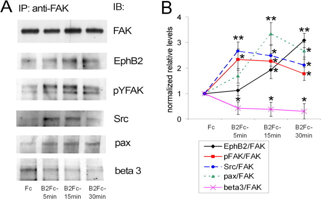
Dendritic spines are the postsynaptic sites of most excitatory synapses in the brain and are highly enriched in polymerized F-actin, which drives the formation and maintenance of mature dendritic spines and synapses. We propose that suppressing the activity of the actin-severing protein cofilin plays an important role in the stabilization of mature dendritic spines, and is accomplished through an EphB receptor-focal adhesion kinase (FAK) pathway. Our studies revealed that Cre-mediated knock-out of loxP-flanked fak prompted the reversion of mature dendritic spines to an immature filopodial-like phenotype in primary hippocampal cultures. The effects of FAK depletion on dendritic spine number, length, and morphology were rescued by the overexpression of the constitutively active FAK(Y397E), but not FAK(Y397F), indicating the significance of FAK activation by phosphorylation on tyrosine 397. Our studies demonstrate that FAK acts downstream of EphB receptors in hippocampal neurons and EphB2-FAK signaling controls the stability of mature dendritic spines by promoting cofilin phosphorylation, thereby inhibiting cofilin activity. While constitutively active nonphosphorylatable cofilin(S3A) induced an immature spine profile, phosphomimetic cofilin(S3D) restored mature spine morphology in neurons with disrupted EphB activity or lacking FAK. Further, we found that EphB-mediated regulation of cofilin activity at least partially depends on the activation of Rho-associated kinase (ROCK) and LIMK-1. These findings indicate that EphB2-mediated dendritic spine stabilization relies, in part, on the ability of FAK to activate the RhoA-ROCK-LIMK-1 pathway, which functions to suppress cofilin activity and inhibit cofilin-mediated dendritic spine remodeling.


Figures

Figure 1.
Figure 1.
Disruption of FAK expression in 14 DIV hippocampal neurons promotes formation of new dendritic protrusions and elongation of existing ones. Neurons were obtained from E15 hippocampi of conditional fak mutant mice bearing loxP-flanked fak alleles, transfected at 12 DIV, and processed for indirect immunofluorescence at 14 DIV. A–C, Confocal images of 14 DIV hippocampal neurons expressing GFP alone (WT) (A), GFP and Cre (FAK KO) (B), or GFP, Cre, and FAK (FAK KO + FAK) (C). Dendritic morphology was observed with GFP fluorescence (green) and FAK expression was detected by immunostaining with anti-FAK antibody (red). Arrows point to neurons with positive FAK immunoreactivity. A double GFP/Cre-expressing neuron with no FAK immunoreactivity is indicated by an arrowhead. Scale bars, 10 μm. D–F, Quantification of dendritic protrusion lengths (D); distribution of dendritic protrusion lengths: <2 μm, 2–4 μm, and >4 μm (E); and dendritic protrusion density in control and FAK-deficient neurons (F). Error bars indicate SEM (n = 200–400 dendritic protrusions from 10 neurons per group; **p < 0.01, ***p < 0.001). FAK-deficient neurons exhibited longer, more numerous filopodia-like protrusions than WT neurons. The effects of FAK depletion on dendritic protrusion number and length were suppressed by FAK overexpression.
Figure 2.
Figure 2.
FAK depletion affects dendritic spine morphology and synapses. A–F, Confocal images of 14 DIV hippocampal neurons expressing GFP alone (WT) (A, D), GFP and Cre (FAK KO) (B, E), or GFP, Cre, and FAK (FAK KO + FAK) (C, F). Neurons were obtained from E15 hippocampi of conditional fak mutant mice bearing loxP-flanked fak alleles, transfected at 12 DIV, and processed for indirect immunofluorescence at 14 DIV. Dendritic spine morphology was observed with GFP fluorescence (green), presynaptic boutons were labeled by immunostaining for the presynaptic vesicle marker synaptophysin (red, A–C), and postsynaptic sites of excitatory synapses were labeled by immunostaining for PSD-95 (red, D–F). Scale bars, 10 μm. G–J, Quantitative analysis of dendritic spine lengths (G), dendritic spine head areas (H), number of synaptophysin- and PSD95-positive puncta (I), and dendritic spine morphology (J). Error bars indicate SEM (n = 200–400 dendritic spines from 10 neurons per group; ***p < 0.001). FAK depletion induces immature dendritic spine profiles in cultured hippocampal neurons. FAK-deficient neurons exhibit more filopodia-like thin spines and less mushroom-shaped dendritic spines. FAK depletion also promotes new filopodia formation, while the number of dendritic spines remains unchanged. These effects of fak deletion on dendritic spine morphology were suppressed by overexpression of FAK.
Figure 3.
Figure 3.
FAK depletion induces actin reorganization in 14 DIV hippocampal neurons. A–C, Confocal images of 14 DIV hippocampal neurons expressing GFP alone (WT) (A), GFP and Cre (FAK KO) (B), or GFP, Cre, and FAK (FAK KO + FAK) (C). Neurons were obtained from E15 hippocampi of conditional fak mutant mice bearing loxP-flanked fak alleles, transfected at 12 DIV, and processed for indirect immunofluorescence at 14 DIV. Dendritic spine morphology was observed with GFP fluorescence (green). Polymerized F-actin was detected in dendritic spines with rhodamine-coupled phalloidin (red). Scale bars, 10 μm. D, E, Quantitative analysis of numbers (D) and sizes (E) of F-actin clusters in control neurons, FAK KO neurons, or FAK KO neurons overexpressing FAK. Data represent the average number of F-actin clusters per 10 μm of dendrite (D) or average size of F-actin clusters (E). Error bars indicate SEM (n = 250–500 F-actin clusters from 10 neurons per group; ***p < 0.001). FAK-deficient neurons show reductions in number and size of F-actin clusters in dendritic spines compared with control neurons. The effects are rescued by overexpression of FAK.
Figure 4.
Figure 4.
FAK activation/phosphorylation and its ability to interact with regulators of Rho family GTPases are required for its effects on dendritic spines. A–F, Confocal images of the dendrites of 14 DIV hippocampal neurons expressing GFP alone (WT) (A), GFP and Cre (FAK KO) (B), GFP, Cre, and FAKY397E (FAK KO + FAKY397E) (C), GFP, Cre, and FAKY397F (FAK KO + FAKY397F) (D), GFP, Cre, and FAKP878A (FAK KO + FAKP878A) (E), or GFP, Cre, and FAKL1034S (FAK KO + FAKL1034S) (F). Neurons were obtained from E15 hippocampi of conditional fak mutant mice bearing loxP-flanked fak alleles, transfected at 12 DIV, and processed for indirect immunofluorescence at 14 DIV. Dendritic spine morphology was observed with GFP fluorescence (green), presynaptic boutons were labeled by immunostaining for the presynaptic vesicle marker synaptophysin (red, A–F). Scale bars, 10 μm. G–I, Quantification of dendritic protrusion lengths (G), dendritic protrusion densities (H), and dendritic spine head area (I) in control and FAK-deficient neurons. Error bars indicate SEM (n = 500–700 dendritic protrusions from 10 neurons per group; ***p < 0.001). Constitutively active FAKY397E, but not inactive FAKY397F mutant, restored mature dendritic spines in FAK-deficient hippocampal neurons.
Figure 5.
Figure 5.
FAK signaling is regulated by EphB receptors. A, 14 DIV hippocampal neurons were treated with ephrin B2-Fc for 5, 15, or 30 min to activate EphB2 receptors. Control cultures were treated with Fc for 15 min. The lysates were immunoprecipitated (IP) with anti-FAK antibody and immunoblotted (IB) with anti-FAK, anti-EphB2, anti-pY397FAK, anti-Src, anti-paxillin, and anti-β3 integrin antibodies. B, The levels of proteins and pY397FAK were quantified by densitometry and normalized to total FAK levels. Experimental values represent mean ± SD (n = 3). Values significantly different in ephrinB2-Fc-treated samples compared with control Fc samples are indicated by asterisks (*p < 0.05; **p < 0.01). EphB2 receptor activation with preclustered ephrin B2-Fc induced long-lasting activation/phosphorylation of FAK and its association with EphB2, Src, and paxillin at 5, 15, and 30 min. Moreover, ephrin B2-induced activation and recruitment of FAK to the EphB2 receptor led to a prolonged dissociation of FAK from β3 integrin, suggesting that the EphB2 receptor may act as a competitive inhibitor of integrin signaling.
Figure 6.
Figure 6.
Constitutively active FAKY397E restores mature dendritic spine morphology disrupted by the inhibition of EphB2 receptor activity. A–C, Confocal images of dendrites of 14 DIV hippocampal neurons expressing GFP alone (GFP) (A), GFP and dnEphB2K662R (GFP + dnEphB2) (B), or GFP, dnEphB2K662R, and FAKY397E (GFP + dnEphB2 + FAKY397E) (C). Neurons were obtained from E15 hippocampi of wt mice, transfected at 12 DIV, and processed for indirect immunofluorescence at 14 DIV. Scale bars, 10 μm. D–G, Quantification of dendritic spine length (D); dendritic spine density (E); distribution of dendritic protrusion lengths: <2 μm, 2–4 μm, and >4 μm (F); and dendritic spine head area (G). Error bars indicate SEM (n = 500 dendritic protrusions from 10 neurons per group; ***p < 0.001). While hippocampal neurons expressing dnEphB2 developed elongated dendritic spines and filopodia, the neurons expressing dnEphB2 together with constitutively active FAKY397E exhibit mature mushroom-like dendritic spines similar to control GFP-transfected neurons.
Figure 7.
Figure 7.
The EphB2 receptor induces cofilin phosphorylation in cultured hippocampal neurons. A, C, Hippocampal neurons (14 DIV) were treated with ephrin B2-Fc to activate EphB receptors or control Fc for 15 min with or without ROCK inhibitor Y27632 at 10 or 75 μm. Cell lysates were subjected to immunoblotting with anti-phospho-cofilin (A) or anti-phospho-LIMK-1/2 (C) antibodies. The blots were stripped and reprobed against total cofilin or LIMK-1. B, The levels of phospho-cofilin were quantified by densitometry and normalized to total cofilin. Experimental values represent mean ± SD (n = 3). Values significantly different in ephrinB2-Fc-treated samples compared with control Fc samples are indicated by asterisks (*p < 0.05; **p < 0.01). Cofilin phosphorylation levels were significantly lower in samples treated with ephrinB2-Fc in presence of 10 μm Y27632 (ap < 0.05) or 75 μm Y27632 (bp < 0.01) than in samples treated with ephrinB2-Fc alone. EphB2 receptor activation with preclustered ephrin B2-Fc led to increased levels of phosphorylated cofilin that was inhibited with ROCK inhibitor Y27632.
Figure 8.
Figure 8.
FAK-mediated cofilin phosphorylation depends on FAK activation/phosphorylation and its ability to interact with regulators of Rho family GTPases. A–E, Confocal images of 14 DIV hippocampal neurons expressing GFP alone (A), GFP and FAK (GFP + FAK) (B), GFP and FAKY397E (GFP + FAKY397E) (C), GFP and FAKY397F (GFP + FAKY397F) (D), or GFP and FAKL1034S (GFP + FAKL1034S) (E). Neurons were obtained from E15 hippocampi of wt mice, transfected at 12 DIV, and processed for indirect immunofluorescence at 14 DIV. Dendritic spine morphology was observed with GFP fluorescence (green), and the distribution of phospho-cofilin (red) and total cofilin (blue) was detected by immunostaining. Scale bar, 10 μm. F, G, Quantification of phospho-cofilin levels (F) and the cofilin/phospho-cofilin ratio (G) in dendrites of transfected (GFP-positive) and control (GFP-negative) neurons. The levels of phospho-cofilin and cofilin were quantified by densitometry. Experimental values represent mean ± SEM (n = 20 dendrites from 5 neurons per group; *p < 0.05; **p < 0.01; ***p < 0.001). H–L, Quantification of dendritic spine length (H); dendritic spine density (I); distribution of dendritic protrusion lengths: <2 μm, 2–4 μm, and >4 μm (J); dendritic spine head area (K); and spine head-area-to-length ratio (L). Error bars indicate SEM (n = 500 dendritic protrusions from 5 neurons per group; *p < 0.05; **p < 0.01; ***p < 0.001).
Figure 9.
Figure 9.
The constitutively active cofilinS3A mutant, but not the inactive cofilinS3D mutant or wt-cofilin, induces immature dendritic spines. A–D, Confocal images of 14 DIV hippocampal neurons expressing dsRed alone (A), dsRed with GFP-wt-cofilin (B), dsRed with GFP-cofilinS3D (C), or dsRed with GFP-cofilinS3A (D). Scale bar, 10 μm. E–H, Quantification of dendritic protrusion length (E), dendritic protrusion density (F), dendritic spine head area (G), and spine head-to-length ratio (H). Error bars indicate SEM (n = 300 dendritic protrusions from 7–10 neurons per group); **p < 0.01; ***p < 0.001. The overexpression of the constitutively active nonphosphorylatable cofilinS3A mutant, but not the dominant-negative phosphomimetic cofilinS3D mutant or wt-cofilin, prompted the reversion of mushroom-shaped mature spines into thin spines with small heads.
Figure 10.
Figure 10.
Cofilin inactivation restores mature dendritic spines in hippocampal neurons with inhibited EphB receptor activity or Cre-mediated fak KO. A–D, I–L, Confocal images of 14 DIV hippocampal neurons expressing dsRed alone (dsRed) (A); dsRed and dnEphB2K662R (dsRed/dnEphB2) (B); dsRed, dnEphB2K662R, and GFP-cofilinS3A (dsRed/dnEphB2/cofilinS3A) (C); dsRed, dnEphB2K662R, and GFP-cofilinS3D (dsRed/dnEphB2/cofilinS3D) (D); dsRed alone (dsRed) (I); dsRed and Cre (dsRed/Cre) (J); dsRed, Cre, and GFP-cofilinS3A (dsRed/Cre/CofilinS3A) (K); or dsRed, Cre, and GFP-cofilinS3D (dsRed/Cre/CofilinS3D) (L). Scale bar, 10 μm. E–H, M–P, Quantification of average dendritic protrusion length (E, M); dendritic protrusion density (F, N); distribution of dendritic protrusion lengths: <2 μm, 2–4 μm, and >4 μm (G, O); and dendritic spine head area (H, P). Error bars indicate SEM (n = 500–800 dendritic protrusions from 7–10 neurons per group); **p < 0.05; ***p < 0.001. The overexpression of the phosphomimetic cofilinS3D mutant, but not the nonphosphorylatable constitutively active cofilinS3A mutant, reversed the effects of EphB2 receptor inactivation and Cre-mediated deletion of fak on dendritic spine morphology.

Comment in

Similar articles

Cited by

References

    1. Abbi S, Guan JL. Focal adhesion kinase: protein interactions and cellular functions. Histol Histopathol. 2002;17:1163–1171. - PubMed
    1. Andrianantoandro E, Pollard TD. Mechanism of actin filament turnover by severing and nucleation at different concentrations of ADF/cofilin. Mol Cell. 2006;24:13–23. - PubMed
    1. Arber S, Barbayannis FA, Hanser H, Schneider C, Stanyon CA, Bernard O, Caroni P. Regulation of actin dynamics through phosphorylation of cofilin by LIM-kinase. Nature. 1998;393:805–809. - PubMed
    1. Asrar S, Meng Y, Zhou Z, Todorovski Z, Huang WW, Jia Z. Regulation of hippocampal long-term potentiation by p21-activated protein kinase 1 (PAK1) Neuropharmacology. 2009;56:73–80. - PubMed
    1. Bamburg JR. Proteins of the ADF/cofilin family: essential regulators of actin dynamics. Annu Rev Cell Dev Biol. 1999;15:185–230. - PubMed

Publication types

MeSH terms