. 2009 Aug;20(16):3660-70.
doi: 10.1091/mbc.e09-01-0009. Epub 2009 Jun 24.

Interaction of CDK5RAP2 with EB1 to track growing microtubule tips and to regulate microtubule dynamics

Affiliations

Interaction of CDK5RAP2 with EB1 to track growing microtubule tips and to regulate microtubule dynamics

Ka-Wing Fong et al. Mol Biol Cell. 2009 Aug.

Abstract

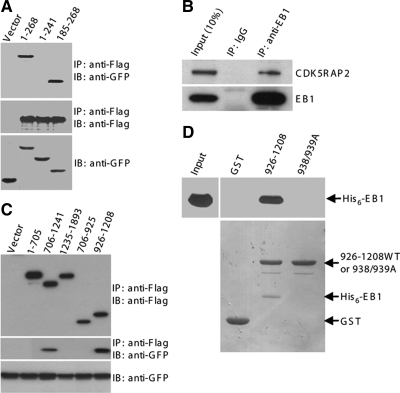
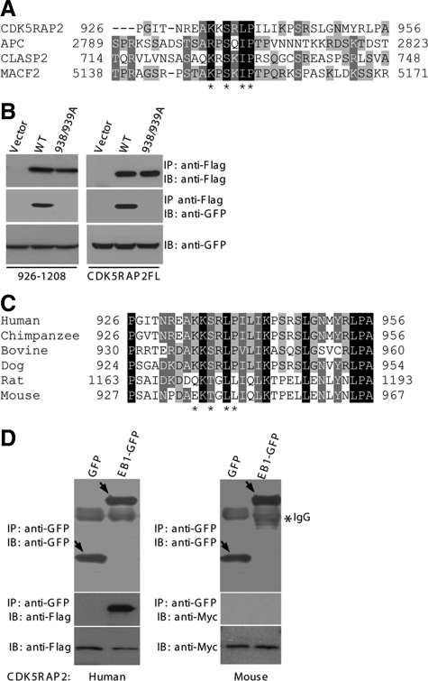
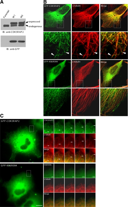
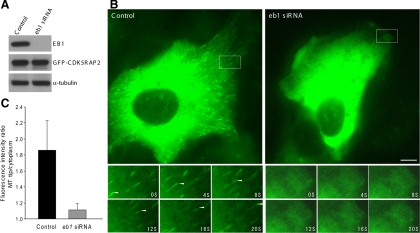
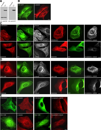
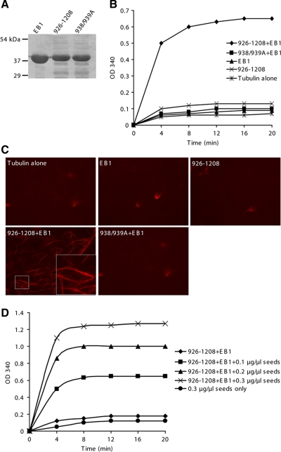
Mutations in cdk5rap2 are linked to autosomal recessive primary microcephaly, and attention has been paid to its function at centrosomes. In this report, we demonstrate that CDK5RAP2 localizes to microtubules and concentrates at the distal tips in addition to centrosomal localization. CDK5RAP2 interacts directly with EB1, a prototypic member of microtubule plus-end tracking proteins, and contains the basic and Ser-rich motif responsible for EB1 binding. The EB1-binding motif is conserved in the CDK5RAP2 sequences of chimpanzee, bovine, and dog but not in those of rat and mouse, suggesting a function gained during the evolution of mammals. The mutation of the Ile/Leu-Pro dipeptide within the motif abolishes EB1 interaction and plus-end attachment. In agreement with the mutational analysis, suppression of EB1 expression inhibits microtubule tip-tracking of CDK5RAP2. We have also found that the CDK5RAP2-EB1 complex regulates microtubule dynamics and stability. CDK5RAP2 depletion by RNA interference impacts the dynamic behaviors of microtubules. The CDK5RAP2-EB1 complex induces microtubule bundling and acetylation when expressed in cell cultures and stimulates microtubule assembly and bundle formation in vitro. Collectively, these results show that CDK5RAP2 targets growing microtubule tips in association with EB1 to regulate microtubule dynamics.


Figures

Figure 1.
Figure 1.
CDK5RAP2 localization to MTs and the distal tips. (A) HeLa cells were stained with anti-CDK5RAP2 and anti-α-tubulin antibodies for confocal microscopy. The boxed areas are enlarged in insets. (B) HeLa cells depleted of CDK5RAP2 by RNAi were immunostained as described in A. (C) The CDK5RAP2 antibody was blocked with antigen protein and then subjected to immunostaining of HeLa cells. Bars, 10 μm.
Figure 2.
Figure 2.
CDK5RAP2 binds to EB1. (A) HEK293T extracts coexpressing Flag-CDK5RAP2 and either GFP or GFP-tagged EB1 proteins were subjected to anti-FLAG immunoprecipitation (IP). The immunoprecipitates (50%) and the cell extracts (5%) were analyzed by immunoblotting (IB). EB1 constructs used were the full-length protein (i.e., 1–268) and two fragments. (B) Endogenous EB1 was immunoprecipitated from HEK293T extracts to detect coimmunoprecipitation of endogenous CDK5RAP2. The immunoprecipitates (100%) and the cell extract (10%) were analyzed. (C) CDK5RAP2 fragments (FLAG-tagged) were coexpressed with EB1-GFP. The anti-FLAG immunoprecipitates (50%) and the cell extracts (5%) were probed on immunoblots. (D) In a GST pull-down assay, His6-EB1 was tested for binding to GST or the CDK5RAP2 fragment in fusion with GST. 938/939A, 926-1208(L938A/P939A). The pull-downs (20%) and the His6-EB1 input (10%) were resolved by SDS-PAGE and transferred to membranes. Top, anti-His6 immunoblots. Bottom, membrane stained with Ponseau S.
Figure 3.
Figure 3.
CDK5RAP2 contains the basic and Ser-rich motif. (A) Alignment of CDK5RAP2(926-956) with several known basic and Ser-rich sequences. Asterisks denote residues conserved in the motif. (B) HEK293T was double transfected with FLAG-tagged CDK5RAP2 and EB1-GFP for anti-FLAG immunoprecipitation. The immunoprecipitates (50%) and the cell extracts (5%) were analyzed on immunoblots. The CDK5RAP2 constructs used were the full-length (CDK5RAP2FL) and the 926-1208 fragment. WT, wild type; 938/939A, L938A/P939A mutant. (C) Sequence alignment of CDK5RAP2 from different mammalian species. GenBank accession numbers are as follows: chimpanzee, NP_001035901; bovine, XP_584826; dog, XP_855524; rat, XP_575844; and mouse, NP_666102. (D) HEK293T extracts coexpressing EB1 and CDK5RAP2 were subjected to anti-GFP immunoprecipitation. The immunoprecipitates (100%) and the cell extracts (5%) were analyzed. CDK5RAP2 constructs are the human protein (FLAG-tagged) and the mouse counterpart (Myc-tagged).
Figure 4.
Figure 4.
CDK5RAP2 targets to MT distal ends in a manner dependant on EB1 interaction. (A) Stable U2OS lines were analyzed on immunoblots (IBs). Parental, parental U2OS; W1, a subline expressing GFP-CDK5RAP2; M3, a subline expressing the L938A/P939A mutant (GFP-938/939A). (B) The stable lines were immunostained for MTs (anti-α-tubulin). (C) Time-lapse microscopy was performed on the stable cells. mCherry-α-tubulin was transiently expressed at low levels. Time series of the boxed areas are enlarged. Bars, 10 μm.
Figure 5.
Figure 5.
Depletion of EB1 inhibits CDK5RAP2 tracking MT plus ends. (A) The stable line of GFP-CDK5RAP2 (clone W1) was transfected with an eb1-targeting siRNA or the control. The protein levels of EB1, GFP-CDK5RAP2, and α-tubulin were detected. (B) Live cells of W1 were imaged for GFP-CDK5RAP2. Below are time series of the boxed areas. Bar, 10 μm. (C) Histogram shows the fluorescence intensity ratios of growing MT tips to cytoplasm. Eight control and twelve EB1-depleted cells were analyzed; 10 MTs were chosen from each cell for analysis. Statistical analysis was performed using Student's unpaired two tails t test (p < 0.001).
Figure 6.
Figure 6.
The CDK5RAP2–EB1 complex induces MT bundling and acetylation. (A) The stable line of EB1-GFP (clone E2) and parental U2OS cells (Parental) were analyzed on immunoblots (IBs). (B) The stable line E2 was imaged for EB1-GFP and MTs (anti-α-tubulin). (C and D) The E2 line was transiently transfected with the wild type or the L938A/P939A mutant (938/939A) of FLAG-CDK5RAP2. The constructs transfected were the full-length protein (C) or the fragment 926-1208 (D). The cells were double stained for CDK5RAP2 (anti-FLAG) and MTs (anti-α-tubulin) or acetylated MTs (anti-acetylated α-tubulin). (E) CDK5RAP2 and 926-1208 were transiently expressed in parental U2OS. The cells were stained as above. Bars, 10 μm.
Figure 7.
Figure 7.
The CDK5RAP2–EB1 complex promotes MT polymerization. (A) The purified recombinant proteins were examined on a SDS-PAGE gel stained with Coomassie Blue. 938/939A, the L938A/P939A mutant of CDK5RAP2(926-1208). (B) MT assembly was performed in the presence of 0.1 μg/μl MT seeds. Recombinant proteins used included 926-1208 or 938/939A, 1.5 μM; and His6-EB1, 0.5 μM. (C) MTs were polymerized as specified in B from a mixture of rhodamine-labeled and unlabeled tubulin for examination by fluorescence microscopy. (D) MT assembly assays contained various amounts of MT seeds. CDK5RAP2(926-1208), 1.5 μM; His6-EB1, 0.5 μM.

References

    1. Akhmanova A., Steinmetz M. O. Tracking the ends: a dynamic protein network controls the fate of microtubule tips. Nat. Rev. Mol. Cell Biol. 2008;9:309–322. - PubMed
    1. Andersen J. S., Wilkinson C. J., Mayor T., Mortensen P., Nigg E. A., Mann M. Proteomic characterization of the human centrosome by protein correlation profiling. Nature. 2003;426:570–574. - PubMed
    1. Askham J. M., Moncur P., Markham A. F., Morrison E. E. Regulation and function of the interaction between the APC tumour suppressor protein and EB1. Oncogene. 2000;19:1950–1958. - PubMed
    1. Askham J. M., Vaughan K. T., Goodson H. V., Morrison E. E. Evidence that an interaction between EB1 and p150(Glued) is required for the formation and maintenance of a radial microtubule array anchored at the centrosome. Mol. Biol. Cell. 2002;13:3627–3645. - PMC - PubMed
    1. Bieling P., Kandels-Lewis S., Telley I. A., van D. J., Janke C., Surrey T. CLIP-170 tracks growing microtubule ends by dynamically recognizing composite EB1/tubulin-binding sites. J. Cell Biol. 2008;183:1223–1233. - PMC - PubMed

Publication types

MeSH terms

Substances