. 2009 Jul 15;183(2):1044-54.
doi: 10.4049/jimmunol.0900420. Epub 2009 Jun 24.

Trans-presentation of IL-15 by intestinal epithelial cells drives development of CD8alphaalpha IELs

Affiliations

Trans-presentation of IL-15 by intestinal epithelial cells drives development of CD8alphaalpha IELs

Lisa J Ma et al. J Immunol. .

Abstract

IL-15 is crucial for the development of intestinal intraepithelial lymphocytes (IEL) and delivery is mediated by a unique mechanism known as trans-presentation. Parenchymal cells have a major role in the trans-presentation of IL-15 to IELs, but the specific identity of this cell type is unknown. To investigate whether the intestinal epithelial cells (IEC) are the parenchymal cell type involved, a mouse model that expresses IL-15Ralpha exclusively by the IECs (Villin/IL-15Ralpha Tg) was generated. Exclusive expression of IL-15Ralpha by the IECs restored all the deficiencies in the CD8alphaalpha(+)TCRalphabeta(+)and CD8alphaalpha(+)TCRgammadelta(+) subsets that exist in the absence of IL-15Ralpha. Interestingly, most of the IEL recovery was due to the preferential increase in Thy1(low) IELs, which compose a majority of the IEL population. The differentiation of Thy1(high)CD4(-)CD8(-) thymocytes into Thy1(-)CD8alphaalpha IELs was found to require IL-15Ralpha expression specifically by IECs and thus, provides evidence that differentiation of Thy1(low) IELs is one function of trans-presentation of IL-15 in the intestines. In addition to effects in IEL differentiation, trans-presentation of IL-15 by IECs also resulted in an increase in IEL numbers that was accompanied by increases in Bcl-2, but not proliferation. Collectively, this study demonstrates that trans-presentation of IL-15 by IECs alone is completely sufficient to direct the IL-15-mediated development of CD8alphaalpha(+) T cell populations within the IEL compartment, which now includes a newly identified role of IL-15 in the differentiation of Thy1(low) IELs.


Figures

Figure 1
Figure 1. IL-15Rα and GFP are expressed in the intestinal epithelium of Villin/IL-15Rα Tg mice
(A) IL-15Rα (red) and GFP (green) was detected in 6 μm sections of small intestine of Villin/IL-15Rα Tg mice. Top row shows IL-15Rα expression as detected using anti-IL-15Rα Ab-biotin followed by visualization with streptavidin-Cy5 while bottom row shows control staining using goat Ig biotin. (B) Histograms in left panel show GFP expression by IECs isolated from Wt and Villin/IL-15Rα Tg mice. Histograms in right panel show cell surface IL-15Rα expression by IECs isolated from the indicated mice. Both panels show fluorescence after gating on CD45-negative cells (IECs). (C) Western blot analysis. Protein from intestinal IECs from Wt, IL-15Rα−/−, and Villin/IL-15Rα Tg mice were analyzed by western blot using anti-IL-15Rα Ab. (D) Phenotype of T cells isolated from spleen, mesenteric lymph node (MLN), and peripheral lymph nodes (PLN). Cells were isolated from the indicated mice and stained for cell surface markers to identify proportions in T cell populations. Left panel shows representative dot plots of lymphocytes stained with CD4 and CD8 from the indicated tissues. Right panel shows staining of CD44 after gating on CD8+ cells; marker designates the proportion of memory-phenotype (CD44hi) cells. (E) Proportion of peripheral NK cells. Lymphocytes from PLN, MLN, and spleen from the indicated mice were stained with NK1.1 and CD3. Box denotes the NK cell population as defined by CD3NK1.1+ staining and numbers represent the average percent of NK cells present.
Figure 2
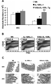
Figure 2. IEC expression of IL-15Rα increases lymphocyte numbers in the intestinal epithelium
(A) Total cell numbers (IELs and IECs) were counted using a hemocytometer and trypan blue exclusion. Total IEL and IEC numbers were determined from the % CD45+ or % CD45neg, respectively. Average of IEL and IEC cell numbers from 4 independent experiments ± SE. *, p<0.05 compared to Wt and Villin/IL-15Rα Tg mice ; **, p<0.001 compared to Wt and IL-15Rα−/− mice. (B) Percent of CD45+ cells among all cells isolated from intestinal epithelium (average of 4 independent experiments ± SE). (C) 6 μm sections of ileum stained for hemotoxylin and eosin.
Figure 3
Figure 3. IL-15-mediated IEL development is recovered in Villin/IL-15Rα Tg mice
(A) Flow cytometric analysis of TCRγδ+ IEL subsets. Top panel shows proportion of CD8α+TCRγδ+ cells among CD45+ IELs. Bottom panel shows staining for Thy1 and Vγ5 expression after gating on CD8α+TCRγδ+ cells. (B) Absolute numbers of the subpopulations of TCRγδ+ IELs isolated from each group of mice (average of 4 independent experiments, n=10−12 mice/group). Circles in top row indicate gating of analysis in bottom row. Error bars represent SE. (C) Flow cytometric analysis of TCRαβ+ IEL subsets. Top panel shows proportion of CD8α+TCRαβ+ cells among CD45+ IELs. Bottom panel shows staining for Thy1 and CD8β expression after gating on CD8α+TCRαβ+ cells. (D) Absolute numbers of the subpopulations of TCRαβ+ IELs isolated from each group of mice (average of 4 independent experiments, n=10−12 mice/group). Error bars represent SE. (E) Flow cytometric analysis of IELs isolated from the colon. First row shows proportion of TCRαβ+ and TCRγδ+ IELs among CD45+ IELs. Remaining rows have gated populations indicated on the left. (F,G) Absolute numbers of (F) TCRγδ+ and (G) TCRαβ+ IELs from the colon. Error bars represent S.D. * p<0.05; ** p<0.01; *** p<0.001 compared to Wt and IL-15Rα−/− mice.
Figure 4
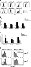
Figure 4. Effects of IL-15Rα expression on Bcl-2 expression and basal proliferation in IEL subsets
(A) Bcl-2 expression measured by intracellular staining after gating on the indicated CD8α+TCRγδ+ IEL subsets (top row) or CD8α+TCRαβ+ IEL subsets (bottom row) isolated from indicated mice. Data are representative of 2 independent experiments (n = 2 mice/group/experiment). (B,C) Mice (6 wk old) were given 2 mg of BrdU i.p. followed by analysis of BrdU incorporation 12 hours later. (B) Average percent BrdU incorporated by TCRγδ IEL subsets. (C) Average percent BrdU incorporated by TCRαβ IEL subsets. (D) The expression of IL-2/15Rβ chain was examined by staining with anti-CD122 Ab on IELs isolated from Wt (bold outline), Villin/IL-15Rα Tg mice (shaded histogram), or IL-15Rα −/− mice (thin outline). Data are representative of 2 independent experiments (n = 2 mice/group/experiment).
Figure 5
Figure 5. In Vivo Differentiation of Thymic IEL Precursors
(A,B) CD4CD8αThy1hi thymocytes were isolated from Wt mice (CD45.1+) and injected i.v. into sub-lethally irradiated (750 cGy) Villin/IL-15Rα Tg, Wt, and IL-15Rα−/− mice (CD45.2+). Recipient mice were analyzed 2−3 weeks later. Flow cytometric plots show phenotype of donor-derived cells among IELs (A) and in spleen (B) from the indicated mice. Gated populations are indicated on the left. (C) CD8αα+Thy1hi or (D) CD8αα+Thy1lo cells were sorted from the IELs of Wt mice (CD45.1+) and transferred into sub-lethally irradiated Villin/IL-15Rα Tg, Wt, and IL-15Rα−/− mice (all CD45.2+). Donor-derived cells present in the intestinal epithelium were analyzed for Thy1 expression 2−3 weeks after transfer. Data are representative of three experiments.

References

    1. Cheroutre H, Lambolez F. The thymus chapter in the life of gut-specific intra epithelial lymphocytes. Curr. Opin. Immunol. 2008;20:185–191. - PMC - PubMed
    1. Lefrancois L. Phenotypic complexity of intraepithelial lymphocytes of the small intestine. J Immunol. 1991;147:1746–1751. - PubMed
    1. Kim SK, Schluns KS, Lefrancois L. Induction and visualization of mucosal memory CD8 T cells following systemic virus infection. Journal of Immunology. 1999;163:4125–4132. - PubMed
    1. Lefrancois L, Parker CM, Olson S, Muller W, Wagner N, Schon MP, Puddington L. The role of beta7 integrins in CD8 T cell trafficking during an antiviral immune response. J Exp. Med. 1999;189:1631–1638. - PMC - PubMed
    1. Jarry A, Cerf-Bensussan N, Brousse N, Selz F, Guy-Grand D. Subsets of CD3+ (T cell receptor alpha/beta or gamma/delta) and CD3- lymphocytes isolated from normal human gut epithelium display phenotypical features different from their counterparts in peripheral blood. Eur. J. Immunol. 1990;20:1097–1103. - PubMed

Publication types