. 2009 Oct;81(4):739-48.
doi: 10.1095/biolreprod.109.076497. Epub 2009 Jun 24.

Linker histones stimulate HSPA2 ATPase activity through NASP binding and inhibit CDC2/Cyclin B1 complex formation during meiosis in the mouse

Affiliations

Linker histones stimulate HSPA2 ATPase activity through NASP binding and inhibit CDC2/Cyclin B1 complex formation during meiosis in the mouse

Oleg M Alekseev et al. Biol Reprod. 2009 Oct.

Abstract

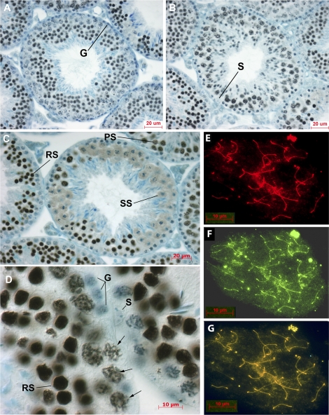
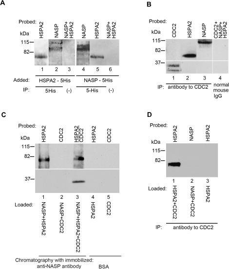
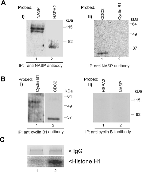
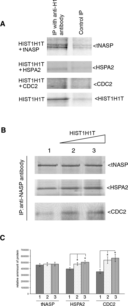
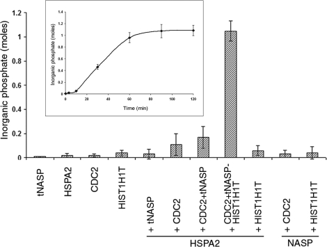
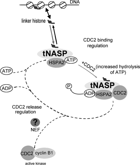
In mammalian spermatocytes, cell division cycle protein 2 (CDC2)/cyclin B1 and the chaperone heat shock protein A2 (HSPA2) are required for the G2-->M transition in prophase I. Here, we demonstrate that in primary spermatocytes, linker histone chaperone testis/embryo form of nuclear autoantigenic sperm protein (tNASP) binds the heat shock protein HSPA2, which localizes on the synaptonemal complex of spermatocytes. Significantly, the tNASP-HSPA2 complex binds linker histones and CDC2, forming a larger complex. We demonstrate that increasing amounts of tNASP favor tNASP-HSPA2-CDC2 complex formation. Binding of linker histones to tNASP significantly increases HSPA2 ATPase activity and the capacity of tNASP to bind HSPA2 and CDC2, precluding CDC2/cyclin B1 complex formation and, consequently, decreasing CDC2/cyclin B1 kinase activity. Linker histone binding to NASP controls the ability of HSPA2 to activate CDC2 for CDC2/cyclin B1 complex formation; therefore, tNASP's role is to provide the functional link between linker histones and cell cycle progression during meiosis.


Figures

FIG. 1.
FIG. 1.
Localization of tNASP in mouse seminiferous tubules and on meiotic chromosomes. A and B) Sections through tubules at stage VI (A) and stage IX (B) show tNASP in leptotene, pachytene, and spermatid stages. Spermatogonia (G) and Sertoli cells (S) do not stain for tNASP. C) A stage XII tubule with diplotene and M-phase spermatocytes proceeding through meiosis to become secondary spermatocytes (SS). Round spermatids (RS) and pachytene spermatocytes (PS) can be seen staining for tNASP in adjacent tubules. D) Higher-magnification image of a seminiferous tubule shows tNASP localized in round spermatid nuclei (RS) and on chromatin in pachytene spermatocytes (arrows). Sertoli cell (S) and spermatogonia (G) nuclei are negative. Control staining with anti-NASP antibody absorbed with antigen was negative (data not shown). EG) Surface-spread meiotic chromosomes demonstrate the localization of tNASP and HSPA2 in mouse primary spermatocytes. E) Staining for tNASP. F) Staining for HSPA2. G) Double staining for tNASP and HSPA2 on chromosomal spreads of mouse spermatogenic cells. Yellow/orange staining demonstrates the colocalization of tNASP and HSPA2. Bars = 20 μm (AC) and 10 μm (DG).
FIG. 2.
FIG. 2.
tNASP-HSPA2 binding in the nuclear fraction of mouse spermatogenic cells. A) Lanes 1 and 2: HSPA2-5His was added to a mouse germ cell nuclear fraction and immunoprecipitated (IP) with anti-5His antibody. Both recombinant HSPA2 (lane 1) and endogenous (nonrecombinant) tNASP (lane 2) were precipitated. In control (lane 3) without anti-5His antibody, neither HSPA2 nor tNASP was immunoprecipitated. Lanes 4 and 5: tNASP-5His was added to a mouse germ cell nuclear fraction and immunoprecipitated with anti-5His antibody. Both recombinant tNASP (lane 4) and endogenous (nonrecombinant) HSPA2 (lane 5) were precipitated. In control (lane 6) without anti-5His antibody, neither HSPA2 nor tNASP was immunoprecipitated. B) Immunoprecipitation of CDC2 (lane 1), HSPA2 (lane 2), and tNASP (lane 3) from the nuclear fraction of mouse spermatogenic cells by anti-CDC2 antibody. Control immunoprecipitation with normal mouse IgG demonstrated that CDC2, tNASP, and HSPA2 (lane 4) did not bind nonspecifically. C) Affinity chromatography with Aminolink-coupled recombinant tNASP bound recombinant HSPA2 (lane 1) but not recombinant CDC2 (lane 2). When HSPA2 and CDC2 were preincubated before addition to the tNASP affinity column, the complex was bound and eluted (lane 3). Control affinity chromatography with Aminolink-coupled bovine serum albumin demonstrated that recombinant HSPA2 (lane 4) and recombinant CDC2 (lane 5) did not bind nonspecifically to Aminolink. D) Anti-CDC2 antibody immunoprecipitates recombinant HSPA2 premixed with recombinant CDC2 (lane 1); however, it does not immunoprecipitate recombinant tNASP premixed with recombinant CDC2 (lane 2). The control (lane 3) shows that anti-CDC2 does not immunoprecipitate recombinant HSPA2.
FIG. 3.
FIG. 3.
A) The presence of increasing amounts of tNASP decreases binding between CDC2 and cyclin B1 in nuclear fractions from mouse spermatogenic cells. Eluates from a CDC2 affinity column loaded with a nuclear fraction from mouse spermatogenic cells only (lane 1) or a nuclear fraction preincubated with increasing amounts of recombinant tNASP at 5 μM (lane 2), 10 μM (lane 3), and 25 μM (lane 4) show a decrease in cyclin B1 binding. The control using affinity beads without CDC2 cross-linked shows no cyclin B1 binding (lane 5). B and C) PhosphorImager detection of 32P-labeled histone indicating that CDC2/cyclin B1 kinase activity decreases in the presence of tNASP and H1 histones. B) tNASP (lane 2), HIST1H1T (lane 3), or tNASP + HIST1H1T (lane 4) was added to mouse testis lysates, and CDC2/cyclin B1 complexes were immunoprecipitated using anti-cyclin B1 antibody. After immunoprecipitation, the kinase activity was determined as a measure of the presence of 32P-labeled histone. The control (lane 1) consisted of a testis lysate alone. *Significant differences in the kinase activity: control vs. tNASP (P < 0.01); control vs. tNASP-HIST1H1T (P < 0.003); and tNASP vs. tNASP-HIST1H1T (P < 0.03). Data are represented as mean ± SD. C) Experiments identical to B were performed using histone HIST1H1A instead of HIST1H1T. Lane 1: control (testis lysate alone). tNASP (lane 2), HIST1H1A (lane 3), or tNASP + HIST1H1A (lane 4) was added to mouse testis lysates, and CDC2/cyclin B1 complexes were immunoprecipitated using anti-cyclin B1 antibody. After immunoprecipitation, the kinase activity was determined as a measure of the presence of 32P-labeled histone. *Significant differences in the kinase activity: control vs. tNASP (P < 0.02); control vs. tNASP-HIST1H1A (P < 0.006); and tNASP vs. tNASP-HIST1H1T (P < 0.05). Data are represented as mean ± SD.
FIG. 4.
FIG. 4.
A) Immunoprecipitation (IP) from the nuclear fraction of mouse spermatogenic cells with anti-NASP antibody precipitated tNASP (I, lane 1), and coimmunoprecipitated HSPA2 (I, lane 2) and CDC2 (II, lane 1) but not cyclin B1 (II, lane 2). B) Immunoprecipitation from the nuclear fraction of mouse spermatogenic cells with anti-cyclin B1 antibody precipitated cyclin B1 (I, lane 1) and CDC2 (I, lane 2) but not HSPA2 (II, lane 1) or tNASP (II, lane 2). C) PhosphorImager detection of 32P-labeled histone indicating specific kinase activity of CDC2 containing immunoprecipitates. Immunoprecipitation with anti-NASP antibody does not have any kinase activity in the precipitate (lane 1); however, when the immunoprecipitation is done with anti-cyclin B1 antibody (lane 2), a strongly radioactive H1 band demonstrates specific kinase activity in the precipitate. Control panel demonstrates equal amounts of antibodies (IgG) used.
FIG. 5.
FIG. 5.
HIST1H1T affects the equilibrium of tNASP-HSPA2-CDC2 complex formation. A) Histone HIST1H1T binds to tNASP but not HSPA2 or CDC2 in vitro. Recombinant HIST1H1T was mixed with recombinant tNASP, HSPA2, CDC2, or HIST1H1T. All lanes were immunoprecipitated (IP) with anti-H1 antibody and stained with their respective antisera (IP with anti-H1). In negative controls (Control IP), HIST1H1T was not added except in the bottom panel (HIST1H1T), where HIST1H1T and a nonrelevant control antibody (anti-CYR61) were added. B) Increasing amounts of HIST1H1T were added to mouse testis lysates and tNASP immunoprecipitated. As HIST1H1T increased, HSPA2 and CDC2 increased: Lane 1: no HIST1H1T was added; lanes 2 and 3: 3 and 6 μM of recombinant HIST1H1T were added, respectively. C) Analysis of bands from B demonstrating significant changes (*) in the quantity of immunoprecipitated HSPA2 and CDC2 in the presence of increasing HIST1H1T. HSPA2: lane 1 vs. lane 2 (P < 0.02), lane 1 vs. lane 3 (P < 0.006). CDC2: lane 1 vs. lane 2 (P < 0.02), lane 1 vs. lane 3 (P < 0.02). The y-axis represents arbitrary units. Data are represented as mean ± SD.
FIG. 6.
FIG. 6.
The HIST1H1T-tNASP-HSPA2-CDC2 complex shows ATPase activity. ATPase activity of HSPA2 is presented as moles of inorganic phosphate liberated by 1 mol of HSPA2 after 60 min of incubation at 37°C in the presence of the indicated proteins. Data are represented as mean ± SD. Inset shows kinetics of HSPA2-mediated ATP hydrolysis in the presence of tNASP, CDC2, and HIST1H1T. Graph presents the moles of inorganic phosphate liberated by 1 mol of HSPA2 during 120 min of incubation at 37°C. Reaction plateau occurs after 60 min of incubation at 37°C.
FIG. 7.
FIG. 7.
Regulation of HSPA2 chaperone cycle dynamics by linker histone-tNASP (modified from Bukau et al. [37] with permission from Elsevier). The cycle regulation starts with the association of linker histones with tNASP and the formation of a linker histone-tNASP-HSPA2 complex. The binding of CDC2 and increased ATP hydrolysis by HSPA2 causes the “locking in” of the substrate (CDC2) and formation of a linker histone-tNASP-HSPA2-CDC2 complex. The predicted release of CDC2 and ADP from the complex would mediate formation of active CDC2/cyclin B1 kinase (dotted line) and depend on nucleotide exchange factors, which are still to be identified.

References

    1. Govin J, Caron C, Lestrat C, Rousseaux S, Khochbin S.The role of histones in chromatin remodelling during mammalian spermiogenesis. Eur J Biochem 2004; 271: 3459–3469.. - PubMed
    1. van der Heijden GW, Derijck AA, Pósfai E, Giele M, Pelczar P, Ramos L, Wansink DG, van der Vlag J, Peters AH, de Boer P.Chromosome-wide nucleosome replacement and H3.3 incorporation during mammalian meiotic sex chromosome inactivation. Nat Genet 2007; 39: 251–258.. - PubMed
    1. Groth A, Rocha W, Verreault A, Almouzni G.Chromatin challenges during DNA replication and repair. Cell 2007; 128: 721–733.. - PubMed
    1. Hochwagen A, Amon A.Checking your breaks: surveillance mechanisms of meiotic recombination. Curr Biol 2006; 16: R217–R228.. - PubMed
    1. Pérez-Hidalgo L, Moreno S, San-Segundo PA.Regulation of meiotic progression by the meiosis-specific checkpoint kinase Mek1 in fission yeast. J Cell Sci 2003; 116: 259–271.. - PubMed

Publication types