. 2009 Sep 24;114(13):2667-77.
doi: 10.1182/blood-2009-02-206532. Epub 2009 Jun 24.

Preclinical characterization of 1-7F9, a novel human anti-KIR receptor therapeutic antibody that augments natural killer-mediated killing of tumor cells

Affiliations

Preclinical characterization of 1-7F9, a novel human anti-KIR receptor therapeutic antibody that augments natural killer-mediated killing of tumor cells

Francois Romagné et al. Blood. .

Abstract

Inhibitory-cell killer immunoglobulin-like receptors (KIR) negatively regulate natural killer (NK) cell-mediated killing of HLA class I-expressing tumors. Lack of KIR-HLA class I interactions has been associated with potent NK-mediated antitumor efficacy and increased survival in acute myeloid leukemia (AML) patients upon haploidentical stem cell transplantation from KIR-mismatched donors. To exploit this pathway pharmacologically, we generated a fully human monoclonal antibody, 1-7F9, which cross-reacts with KIR2DL1, -2, and -3 receptors, and prevents their inhibitory signaling. The 1-7F9 monoclonal antibody augmented NK cell-mediated lysis of HLA-C-expressing tumor cells, including autologous AML blasts, but did not induce killing of normal peripheral blood mononuclear cells, suggesting a therapeutic window for preferential enhancement of NK-cell cytotoxicity against malignant target cells. Administration of 1-7F9 to KIR2DL3-transgenic mice resulted in dose-dependent rejection of HLA-Cw3-positive target cells. In an immunodeficient mouse model in which inoculation of human NK cells alone was unable to protect against lethal, autologous AML, preadministration of 1-7F9 resulted in long-term survival. These data show that 1-7F9 confers specific, stable blockade of KIR, boosting NK-mediated killing of HLA-matched AML blasts in vitro and in vivo, providing a preclinical basis for initiating phase 1 clinical trials with this candidate therapeutic antibody.


Figures

Figure 1
Figure 1
Specificity of 1-7F9 antibody. (A) Characterization of 1-7F9 specificity for KIR2D subtypes. Transduced BWZ cells expressing individual KIR2DL or KIR2DS receptors were incubated for 30 minutes with anti-KIR antibodies (1 μg/mL), as indicated. The 1-7F9 was detected with PE anti–human IgG4, and EB6, GL183, and FES172 were revealed with PE goat anti–mouse IgG. Results shown are representative of 4 separate experiments. (B) Human whole blood from a healthy volunteer was stained with PE-conjugated 1-7F9; dot plots represent 1-7F9 binding to the indicated leukocyte subsets based on forward/side light scatter. (C) Human whole blood from a healthy volunteer was stained with PE-conjugated 1-7F9 and a combination of mAbs defining various leukocyte subsets, and analyzed by flow cytometry. Experiments in panels B and C have been performed on 11 healthy donors. Mean percentage and SD of 1-7F9–positive cells among the NK- and T-cell populations were 48.1% ± 14.9 and 2.4% ± 2.1, respectively. (D) Titration of 1-7F9 mAb on KIR2D-transduced BWZ cell lines. Cells were incubated for 30 minutes with 1/3 serial dilutions of 1-7F9, which were then revealed with PE anti–human IgG4 and analyzed by flow cytometry. Each dilution point was performed in duplicate. formula image, ■, ▲, and ● represent cell lines expressing KIR2DL1, KIR2DS1, KIR2DL3, and KIR2DS2, respectively. Mean and SD of data collected in 2 independent experiments are shown.
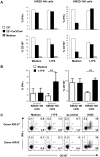
Figure 2
Figure 2
1-7F9 blocks interactions of inhibitory KIR2DL with HLA class I on B-EBV cells. (A) Binding of soluble KIR2DL1-hFc was blocked by anti-KIR mAbs GL183 or DF200 and binding of KIR2DL1-mFc blocked by 1-7F9, as measured by flow cytometry. Relative binding of KIR-Fc proteins to .221-Cw4 cells is shown as percentage of binding by KIR-Fc in the absence of mAbs. Similar data were obtained in a repeat experiment. (B) In a 51Cr release cytotoxicity assay, YTS cells efficiently killed LCL721.221-Cw4 cells (▲), whereas YTS-2DL1 cells did not (●; E:T ratio 12:1). Preincubation (30 minutes at 37°C) of the NK cells with increasing doses of 1-7F9 augmented the killing of LCL721.221-Cw4 targets by YTS-2DL1 cells, in a dose-dependent manner. Curve fitting using one-site receptor saturation equation gives an EC50 of 0.71 μg/mL (95% CI, 0.2-1.2 μg/mL). Experiment shown is representative of multiple experiments giving equivalent results. (C) NK-cell clones were tested for cytolytic activity against B-EBV cell lines transfected with indicated HLA class I allotypes at 10:1 E:T ratio, with or without mAb at 10 μg/mL. NK clone MDC14 (top panels) is KIR2DL1+, KIR2DL2/3, NKG2A. NK clone MDC88 (bottom panels) is KIR2L1, KIR2DL2+, KIR2DL3, NKG2A. Killing by these NK cells was tested against B-EBV cell lines transfected with HLA-Cw4 (left panels) and HAL-Cw1 (right panels). Results were analyzed by ANOVA, followed by Bonferroni posttest; only P < .001 are reported.
Figure 3
Figure 3
1-7F9 interferes with HLA class I-induced inhibitory signaling in KIR2DL-positive resting NK cells. (A) Freshly purified human NK cells were incubated overnight in medium. Then NK cells were incubated for 4 hours at 37°C alone (medium; ▩), with rituximab-coated 221 cells (E:T ratio = 1; □), or rituximab-coated 221-Cw3/Cw4 (E:T ratio = 1; ■) in the presence or not of 1-7F9 (10 μg/mL) and with anti-CD107 and monensin. Cells were then stained with anti-CD3, anti-CD56, and purified 1-7F9 revealed by anti–human IgG4 (HP6025); fixed; permeabilized; and finally stained with anti–IFN-γ. Percentage of IFN-γ positive (top panels) and CD107 positive (bottom panels) was then assessed on KIR2D-negative NK cells (left panels) and KIR2D-positive NK cells (right panels). Results from 1 representative donor are shown. (B) Data represent means ± SD of the percentages of IFN-γ–positive NK cells (left panel) and of CD107-positive NK cells (right panel) collected using method described in panel A, from 6 people. Results in the presence of 221-Cw3/Cw4 are shown. Statistical analysis was performed using first a one-way repeated measures ANOVA test, followed by a Bonferroni test to compare pairs of values (ie, KIR2D medium with KIR2D 1-7F9 and KIR2D+ medium with KIR2D+ 1-7F9). **P < .01. Multiple experiments have also been performed with 221 transfected with single HLA-C and provided similar results. (C) Thawed human PBMC from KIR-S–positive (1 and 2) or KIR-S–negative (3 and 4) donors were incubated for 4 hours at 37°C, alone or in the presence of 1-7F9 (10 μg/mL), cognate isotypic control (IgG4, 10 μg/mL), or K562 (E:T ratio = 10) in the presence of anti-CD107 and monensin. After incubation, cells were stained with anti-CD3 and anti-CD56, and then fixed, permeabilized, and stained with anti–IFN-γ. CD107 mobilization and IFN-γ production are then assessed on NK cells (CD3CD56+ lymphocytes). Results are representative of 1 experiment of 2 done with a KIR-S–positive and a KIR-S–negative donor. Percentage of cells in each quadrant is shown.
Figure 4
Figure 4
1-7F9 increases NK cell–mediated lysis of primary AML blasts. (A) Lysis of primary AML cells by IL-2–activated NK cells of a healthy, KIR ligand-matched donor, in the presence of 1-7F9 at 10 μg/mL (▨) or without antibody (□), measured in standard 51Cr assay at the indicated E:T ratios. Differences were analyzed by Student t test (***P < .005). Experiment shown is representative of different experiments using different effector and target blast cells. (B) Patient NK cells expanded in IL-2 were mixed with freshly thawed, autologous AML blasts obtained at diagnosis. The 1-7F9, control hIgG4, or a mixture of mouse anti-KIR2DL1 and -2/3 F(ab′)2 fragments was added to each well (each at 30 μg/mL final concentration), and after 4 hours, percent specific lysis was measured by a flow cytometry–based assay. Results shown are representative of 4 independent experiments. (C) Patient NK cells (n = 4 for the E:T 30:1 level, plus n = 5 at E:T 15:1) were incubated with 1-7F9 or hIgG4 isotype control mAb and cocultured in the presence of autologous AML blasts. Cytotoxicity, measured as granzyme B release, is shown. Statistically significant differences in cytotoxicity (*P = .01; **P < .03), as a function of granzyme B release were observed at multiple E:T ratios as shown (■ = 1-7F9; ▩ = IgG4 isotype control). No appreciable granzyme B release was found in effectors and targets cultured alone (negative controls for the assay). As a positive control for the ELISPOT granzyme B assay readout, an equivalent number of NK cells from a healthy donor was cultured in 30:1 ratio against the NK cell–sensitive K562 cell line (■). Results are representative of 2 independent experiments in 2 patients.
Figure 5
Figure 5
Receptor occupancy and functional activity of 1-7F9 in KIR2DL3tg mice. (A) KIR2DL3 is functional in tg mice. Splenocytes from wild-type (□) and from genetically modified mice (▩, KbDbKO cells; ■, KbDbKO Cw3tg cells), labeled with 0.5 and 3 μM CFSE, respectively, were mixed 1:1 (10 million each) and injected (i.v.) into wild-type or KIR2DL3tg mice. Twenty hours later, the mean percentage ± SD (3 to 6 mice per group) of cells with each level of CFSE was determined by flow cytometry. Results were analyzed by ANOVA, followed by Bonferroni posttest (***P < .005). (B) Single injection of 1-7F9 induces rejection of HLA-Cw3–positive splenocytes in a NK-dependent manner. Spleen cells from wild-type and KbDbKO, Cw3tg mice were labeled differentially with CFSE, as in panel A, and were injected into Rag KO, KIR2DL3tg mice. Four hours before the cells, 1-7F9 and NK1.1 mAbs were injected intravenously at the indicated doses. Twenty hours after injection of cells, percentage of cells with each level of CFSE was determined in blood (□) and spleen (▩) by flow cytometry, and the ratio of tg to wild-type donor cells is reported. In some animals, NK cells were depleted by administration of NK1.1 mAb (200 μg per mouse, intravenously). Curve fitting using receptor saturation equation gives an EC50 of 4.6 μg per mouse (95% CI, 1.5-7.7). Experiment shown is representative of 2 different experiments. (C) Maximum effect of 1-7F9 is achieved when KIR2DL3 receptor is saturated. The level of KIR2DL3 receptor occupancy after injection of 1-7F9 was estimated by flow cytometry. Cells from blood and spleen of mice were stained ex vivo with PE-conjugated 1-7F9, and the total MFI of the NK population (a measure of the free receptors) is determined. The level of receptor saturation at a given dose is calculated as the ratio of MFI obtained in mice injected with 1-7F9 relative to MFI obtained in untreated mice (3 mice per group). Receptor occupancy was measured in parallel with the determination of ratio of CFSE-labeled cells in B (24 hours after injection of 1-F9).
Figure 6
Figure 6
1-7F9–induced clearance of AML cells by human NK cells in NOD-SCID mice. (A) Flow cytometric analysis of KIR and NKG2A expression by the polyclonal IL-2–activated NK-cell population that was used as effector cells in the in vitro cytotoxicity and the in vivo tumor rejection experiments shown in panels B and C, respectively. (B) □, Lysis of primary human AML cells by KIR ligand-matched NK cells, without (□) or with 1-7F9 antibody (formula image). (The NK cells were isolated from a healthy donor having the same HLA class I allotype groups as the AML target cells.) E:T ratio was 15:1. Results were analyzed by Student t test (***P < .005). (C) NOD-SCID mice infused with autologous NK cells and AML target cells at 1:3 E:T ratio died of leukemia within 65 days. Treatment with 1-7F9 (250 μg/mouse) rescued mice challenged with NK and AML cells at an E:T ratio of 1:12, but not at an E:T of 1:18. N = 5 mice per group. Results have been analyzed by Kaplan Meier log rank test (***P < .005). Similar results were obtained in a repeat experiment.

Comment in

Similar articles

Cited by

References

    1. Vivier E, Tomasello E, Baratin M, Walzer T, Ugolini S. Functions of natural killer cells. Nat Immunol. 2008;9:503–510. - PubMed
    1. Lanier LL. NK cell recognition. Annu Rev Immunol. 2005;23:225–274. - PubMed
    1. Moretta L, Moretta A. Unravelling natural killer cell function: triggering and inhibitory human NK receptors. EMBO J. 2004;23:255–259. - PMC - PubMed
    1. Gasser S, Orsulic S, Brown EJ, Raulet DH. The DNA damage pathway regulates innate immune system ligands of the NKG2D receptor. Nature. 2005;436:1186–1190. - PMC - PubMed
    1. Bottino C, Castriconi R, Moretta L, Moretta A. Cellular ligands of activating NK receptors. Trends Immunol. 2005;26:221–226. - PubMed

Publication types

MeSH terms