. 2009 Sep 4;284(36):24415-24.
doi: 10.1074/jbc.M109.003244. Epub 2009 Jun 24.

A progesterone receptor co-activator (JDP2) mediates activity through interaction with residues in the carboxyl-terminal extension of the DNA binding domain

Affiliations

A progesterone receptor co-activator (JDP2) mediates activity through interaction with residues in the carboxyl-terminal extension of the DNA binding domain

Krista K Hill et al. J Biol Chem. .

Abstract

Progesterone receptor (PR) belongs to the nuclear receptor family of ligand-dependent transcription factors and mediates the major biological effects of progesterone. Transcriptional co-activators that are recruited by PR through the carboxyl-terminal ligand binding domain have been studied extensively. Much less is known about co-activators that interact with other regions of receptors. Jun dimerization protein 2 (JDP2) is a PR co-activator that enhances the transcriptional activity of the amino-terminal domain by increasing the alpha-helical content and stability of the intrinsically disordered amino-terminal domain. To gain insights into the mechanism of JDP2 co-activation of PR, the structural basis of JDP2-PR interaction was analyzed using NMR. The smallest regions of each protein needed for efficient protein interaction were used for NMR and included the basic region plus leucine zipper (bZIP) domain of JDP2 and the core zinc modules of the PR DNA binding domain plus the intrinsically disordered carboxyl-terminal extension (CTE) of the DNA binding domain. Chemical shift changes in PR upon titration with JDP2 revealed that most of the residues involved in binding of JDP2 reside within the CTE. The importance of the CTE for binding JDP2 was confirmed by peptide competition and mutational analyses. Point mutations within CTE sites identified by NMR and a CTE domain swapping experiment also confirmed the functional importance of JDP2 interaction with the CTE for enhancement of PR transcriptional activity. These studies provide insights into the role and functional importance of the CTE for co-activator interactions.


Figures

FIGURE 1.
FIGURE 1.
The basic domain of JDP2 is the minimal region to interact with PR. A, schematic of the nuclear receptor DBD showing the Gly-Met (GM) boundary distinguishing the core DBD from the CTE. Open circles represent amino acids, and light gray circles represent helical regions. Coordinated zinc (Zn) and cysteine (C) residues are labeled. Amino acids comprising the proximal box (P box) have lines drawn through them, whereas residues comprising the dimerization box (D box) are darker gray. B, constructs of PR and JDP2 used in protein-protein interaction assays including PR DBD-CTE641, GST·JDP2, GST·JDP2 amino terminus (N) +basic domain, GST·JDP2 bZIP, GST·JDP2 basic domain, and GST·JDP2 ZIP. C, PR DBD-CTE641 was incubated with free GST or the different GST·JDP2 fusion proteins immobilized to glutathione-Sepharose resins. After washing resins, bound protein was eluted and detected by immunoblot assay with a PR DBD-specific antibody along with input PR DBD-CTE641.
FIGURE 2.
FIGURE 2.
NMR mapping of JDP2 interaction with the PR DBD-CTE. A, HSQC spectrum of PR DBD-CTE641. NMR spectra were recorded at 15 °C on a Varian 600-MHz spectrometer using a triple-resonance (1H/13C/15N) pulsed field gradient cold probe. B, unlabeled JDP2 bZIP was titrated into PR DBD-CTE641, and multiple HSQC experiments were recorded on the Varian 600-MHZ spectrometer. Overlay resonance assignments of PR DBD-CTE641 alone are shown in blue, and PR DBD-CTE641 plus JDP2 bZIP assignments are shown in red. Resonances that shift or disappear on the addition of JDP2 bZIP indicate sites on PR DBD-CTE641 that are perturbed by the binding of JDP2.
FIGURE 3.
FIGURE 3.
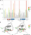
Overlay of NMR chemical shift changes in the presence of JDP2 on the structure of the PR DBD-CTE. A, chemical shift changes between PR DBD-CTE641 alone and PR DBD-CTE641 with JDP2 bZIP were quantitated by [0.5(ΔNH2 + ΔN2/25)]½ (74). Larger differences indicate more of a shift change upon the addition of JDP2 bZIP. Resonances with the largest perturbations by JDP2 are colored in red. D box, dimerization box. B, color-coded map of PR DBD-CTE crystal structure (21) showing areas of chemical shift changes in the presence of JDP2 bZIP; red represents the greatest change, and gray represents the least change.
FIGURE 4.
FIGURE 4.
Specific residues in the CTE are required for JDP2-PR binding. A, single amino acid substitutions were introduced into PR DBD-CTE641, and the effects of the mutations on binding to JDP2 were analyzed by pulldown assay. Varying amounts of WT or mutant PR DBD-CTE641 were incubated with either free GST or GST·JDP2·bZIP immobilized to glutathione-Sepharose resins. Bound protein was eluted after washing of beads and detected along with input PR DBD-CTE641 by immunoblotting with a PR DBD-specific antibody. B, the two amino acid substitutions indicated were introduced into PR DBD-CTE648 and compared with wild type (WT) PR DBD-CTE648 for binding to JDP2·bZIP by GST pulldown as in A above.
FIGURE 5.
FIGURE 5.
A subregion of the CTE is required for JDP2-PR binding. A, schematic of PR DBD-CTE truncations used in GST pulldown assays. B, varying amounts of PR DBD-CTE constructs were incubated with either free GST or GST·JDP2·bZIP immobilized to glutathione-Sepharose resins. Bound protein along with input PR DBD-CTEs was detected by immunoblot with a PR DBD-specific antibody.
FIGURE 6.
FIGURE 6.
The PR CTE alone is sufficient for binding JDP2-PR binding. A, sequences of synthetic peptides used to compete for JDP2-PR binding. The CTE peptide is a 19-mer corresponding to aa 632–656 of the PR CTE. sCTE (scrambled) is a 19-mer containing the same amino acid composition as the CTE except that the sequence was randomly scrambled. B, PR DBD-CTE650 was incubated with either free GST or GST·JDP2·bZIP immobilized to glutathione-Sepharose resins in the presence of varying amounts of either CTE or sCTE peptides. Bound protein along with input PR DBD-CTE650 were eluted after washing resins and detected by immunoblotting with a PR DBD-specific antibody.
FIGURE 7.
FIGURE 7.
Functional requirement of CTE for JDP2 enhancement of PR-mediated gene transcription in cells. A, mutations in CTE enhance progesterone-dependent transcription activity of PR in the absence of ectopically expressed JDP2. Cos-1 cells were transiently co-transfected with a PRE2-TATA-luciferase reporter gene and an expression plasmid for wild type PR-B or PR-B containing substitution mutations in the CTE (R637A/K638A). Cells were treated without (vehicle) or with progesterone (Prog) (10 nm) for 48 h and assayed for luciferase activity as described under “Experimental Procedures.” Results were calculated as relative luciferase activity in vehicle versus progesterone treated cells and are average values ± S.E. (error bars) from three independent experiments. Paired type 1 Student's t test analysis was used to determine statistical significance for the difference of progesterone induction between PR-B and PR-B R637A/K638A; p < 0.05. The inset is an immunoblot of cell extracts used in luciferase assays with a PR-specific antibody (1294) to detect relative levels of WT PR-B and PR-B R637A/K638A mutant protein expression (PR-B is 118 kDa). B, mutations in CTE attenuated JDP2 enhancement of PR transcriptional activity. Cos-1 cells were co-transfected as in A above with PR-B or PR-B CTE (R637A/K638A) and PRE-TATA-luc except in the absence (empty vector) or presence of varying amounts (25, 50, 75 ng) of JDP2 expression plasmids. Results were calculated as -fold JDP2 stimulation of PR-mediated transactivation of luciferase reporter gene activity in the presence of progesterone by setting relative luciferase activity of each receptor in the absence of transfected JDP2 to 1.0. Data represent average values ± S.E. from three independent experiments. Paired type 1 Student's t test analysis was used to determine statistical significance between the effect of JDP2 on PR-B and PR-B R637A/K638A; p < 0.05. C, swapping of TR for PR CTE eliminated JDP2 stimulation of PR transcriptional activity. Cos-1 cells were co-transfected as in B except with PR-B or a PR·TR chimera consisting of a swap within full-length PR-B of the CTE from TR for PR CTE. Results were calculated as the -fold increase of JDP2 stimulation in the presence by setting relative luciferase activity of each receptor in the absence of transfected JDP2 to 1.0. Data represent average values ± S.E. from three independent experiments. Paired type 1 Student's t test analysis was used to determine statistical significance between effects of JDP2 on PR-B and PR·TR chimera; p < 0.05.
FIGURE 8.
FIGURE 8.
Working hypothesis for the CTE as a multidimensional regulatory region of progesterone receptor. The CTE is proposed to be a short, intrinsically disordered region of PR that is capable of dynamic interactions with the minor groove of DNA or with multiple proteins including JDP2 or others such as HMGB. Distinct as well as overlapping residues in the CTE are important for interaction with different partners; aa 632–641 is most important for interaction with DNA and JDP2, whereas aa 642–656 is more important for interaction with HMGB (31). The CTE is capable of adopting distinct conformations dependent on the nature of the interacting partner, and JDP2-induced structural changes in the CTE are proposed to mediate coupling with the disordered NTD of PR.

Similar articles

Cited by

References

    1. Mangelsdorf D. J., Thummel C., Beato M., Herrlich P., Schütz G., Umesono K., Blumberg B., Kastner P., Mark M., Chambon P., Evans R. M. (1995) Cell 83, 835–839 - PMC - PubMed
    1. Chawla A., Repa J. J., Evans R. M., Mangelsdorf D. J. (2001) Science 294, 1866–1870 - PubMed
    1. Li X., O'Malley B. W. (2003) J. Biol. Chem. 278, 39261–39264 - PubMed
    1. Meyer M. E., Quirin-Stricker C., Lerouge T., Bocquel M. T., Gronemeyer H. (1992) J. Biol. Chem. 267, 10882–10887 - PubMed
    1. Bourguet W., Germain P., Gronemeyer H. (2000) Trends Pharmacol. Sci. 21, 381–388 - PubMed

MeSH terms

LinkOut - more resources