. 2009 Jul 30;460(7255):627-31.
doi: 10.1038/nature08209.

Adult satellite cells and embryonic muscle progenitors have distinct genetic requirements

Affiliations

Adult satellite cells and embryonic muscle progenitors have distinct genetic requirements

Christoph Lepper et al. Nature. .

Abstract

Myogenic potential, survival and expansion of mammalian muscle progenitors depend on the myogenic determinants Pax3 and Pax7 embryonically, and Pax7 alone perinatally. Several in vitro studies support the critical role of Pax7 in these functions of adult muscle stem cells (satellite cells), but a formal demonstration has been lacking in vivo. Here we show, through the application of inducible Cre/loxP lineage tracing and conditional gene inactivation to the tibialis anterior muscle regeneration paradigm, that, unexpectedly, when Pax7 is inactivated in adult mice, mutant satellite cells are not compromised in muscle regeneration, they can proliferate and reoccupy the sublaminal satellite niche, and they are able to support further regenerative processes. Dual adult inactivation of Pax3 and Pax7 also results in normal muscle regeneration. Multiple time points of gene inactivation reveal that Pax7 is only required up to the juvenile period when progenitor cells make the transition into quiescence. Furthermore, we demonstrate a cell-intrinsic difference between neonatal progenitor and adult satellite cells in their Pax7-dependency. Our finding of an age-dependent change in the genetic requirement for muscle stem cells cautions against inferring adult stem-cell biology from embryonic studies, and has direct implications for the use of stem cells from hosts of different ages in transplantation-based therapy.


Figures

Figure 1
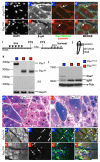
Figure 1. Adult-specific Pax7 mutant cells regenerate muscle efficiently
a, Tmx and CTX regimen and regeneration assay scheme. Vertical lines, daily intervals. b, TA muscle diagram with injury in grey. Horizontal lines, cross sections; W.B., Western blot. c-f, β-gal (by X-galactosidase reaction) and Nuclear Fast Red (NFR) stained Pax7+/CE (c, d) and Pax7CE/f (e, f) muscles at 5 (c, e) and 10 (d, f) days post injury. Asterisks, regenerating fibers with central nuclei; dashed lines, boundary of injury. g, h, Mean diameter (g) and mean inner radius (h) of 10-day regenerated β-gal+ fibers; 300 fibers / animal (n = 3 / genotype); error bars = s.d.; p, two-tailed student's t test. i, j, RT-PCR (i) and W.B. (j) of examples of Pax7+/CE and Pax7CE/f (2 shown) muscles; wild type Pax7 and recombined (Pax7Δ) transcripts as indicated; -RT control, Gapdh control, endogenous IgG1, and α-tubulin (α-TUB, loading control) as indicated. k, l, β-gal and NFR stained Pax7CE/CE muscles at 5 (k) and 10 (l) days post injury; regimen in a. All animals were R26R+/−. Scale bars = 25 μm for c, e in e; for d, f in f; for k, l in l.
Figure 2
Figure 2. Adult-specific Pax7 mutant cells are functional satellite cells
a-h, Fluorescent microscopy of Pax7+/CE (a-d) and Pax7CE/f (e-h) 10-day regenerates: DAPI (a, e); β-gal (b, f); Laminin (red), Pax7 and M-Cad (both in green: Pax7, nuclear, and M-Cad, satellite/muscle junction) (c, g). d and h are merged from a-c and e-g, respectively; β-gal pseudo-colored in red, DAPI in blue, Laminin in cyan, Pax7 and M-Cad in green; white arrows, satellite cells. i, Double injury regeneration assay scheme with EdU injections; labeling as in Fig. 1a with 14 days (14d) between two injuries. j, TA muscle diagram with 1st injury in grey, 2nd injury in black. k, l, RT-PCR (k) and W.B. (l) of TA muscles after second injury; labeling as in Fig. 1i, j. m, n, β-gal and NFR stained Pax7+/CE (m) and Pax7CE/f (n) regenerates at day-6 post 2nd injury; dashed lines, boundaries between 1st and 2nd regenerations based on differences in fiber size; white and black asterisks, old and new regenerated fibers, respectively. All fibers are β-gal+ compared to interstitial cells, but with varying staining signals. o-v, Fluorescent microscopy of Pax7+/CE (o-r) and Pax7CE/f (s-v) regenerates: β-gal (o, s); EdU (p, t); RNMy2/9D2 (green) and DAPI (blue) (q, u). r and v are merged from o-q and s-u, respectively; β-gal in red and other colors same as panels to the left; EdU+ nuclei, white arrows. Scale bars = 25 μm for a-h in h; for m, n in n; for o-r in r; for s-v in v.
Figure 3
Figure 3. Pax3 and Pax7 are dispensable for adult muscle regeneration
a, b, RT-PCR (a) and W.B. (b) of indicated genotypes from regeneration assay scheme in Fig. 1a. Labeling is the same as Fig. 1i, j, with additions of Pax3 wild type and recombined (Pax3Δ) transcripts. c-h, CreEsr+/- ;Pax7+/f (c, e, f) and CreEsR+/−;Pax3/7f/f (d, g, h) day-10 regenerated muscles: hematoxylin and eosin stained (c, d); fluorescent microscopy of Pax7 and M-Cad together (arrowheads, M-Cad+ cells) (e, g); f and h, colored composites with co-stained Laminin (red), DAPI (blue), and Pax7/M-Cad (green) of e and g, respectively; asterisks, central nuclei; arrows, satellite cells. i, j, Mean diameter (i) and mean inner radius (j) of regenerated fibers; 300 fibers / animal (n = 3 / genotype); error bars = s.d.; p, two-tailed student's t test. Scale bars = 25 μm for c, d in d; e-h, in h.
Figure 4
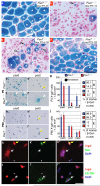
Figure 4. Age-dependent intrinsic change of Pax7 requirement
a-d, Regeneration assessed at day-10 by β-gal and NFR staining: a, Control (Pax7+/CE) regenerates of P7-P11 tmx-treated sample; essentially the same for other time points (not shown). b-d, Conditional inactivation of Pax7 (Pax7CE/f) by tmx at P7-11 (b), P14-18 (c), and P21-25 (d) followed by injury at P21 for (b, c), and P26 for (d); RT-PCR confirmed complete Pax7 inactivation (not shown); new fibers, asterisks; triangles, β-gal+ cells; n = 3 each; all are R26R+/−. e-j, P0 (e-g) and adult (P60, h-j) tmx-treated (0.4 μM) and β-gal stained Pax7+/CE;R26R+/− (e, e′, h, h′) and Pax7CE/f;R26R+/− (f, f′, i, i′) myoblasts at passages 0 (pas0, e, f, h, i) and 2 (pas2, e′, f′, h′, i′); yellow arrows, round/short-spindled myoblasts; white arrows, fibroblastic cells. g, j, Left, average cell numbers (≥ 9 fields) at each passage (pas, x-axes); n = 3 /genotype /stage; error bars = s. d.; right, percentages of Desmin+ (D) and ER-TR7+ (E) of β-gal+ cells at passage 1 (n ≥ 200 each). Fluorescent microscopy of tmx-treated P0 Pax7CE/f;R26R+/− myoblasts at passage 1. k, l, β-gal (k, l), Desmin (k′), ER-TR7 (l′), and merged with DAPI (k″ with k, k′; l″ with l, l′); yellow arrows, Desmin+ cells; white arrows, ER-TR7+ or fibroblastic cells; white dots, negative cells for reference; note β-gal staining and immunofluorescence in nuclei. Scale bars = 50 μm for a-d in d, for e-i′ in i′, and for k-l″ in to l″.

Comment in

References

    1. Relaix F, Rocancourt D, Mansouri A, Buckingham MA. Pax3/Pax7-dependent population of skeletal muscle progenitor cells. Nature. 2005;435:948–953. - PubMed
    1. Seale P, et al. Pax7 is required for the specification of myogenic satellite cells. Cell. 2000;102:777–786. - PubMed
    1. Oustanina S, Hause G, Braun T. Pax7 directs postnatal renewal and propagation of myogenic satellite cells but not their specification. Embo J. 2004;23:3430–3439. - PMC - PubMed
    1. Kuang S, Charge SB, Seale P, Huh M, Rudnicki MA. Distinct roles for Pax7 and Pax3 in adult regenerative myogenesis. J Cell Biol. 2006;172:103–113. - PMC - PubMed
    1. Relaix F, et al. Pax3 and Pax7 have distinct and overlapping functions in adult muscle progenitor cells. J Cell Biol. 2006;172:91–102. - PMC - PubMed

Publication types

MeSH terms