Giant lipomas of the upper extremity
- PMID: 19554145
- PMCID: PMC2687496
- DOI: 10.1177/229255030701500308
Giant lipomas of the upper extremity
Abstract
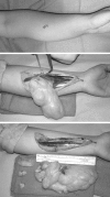
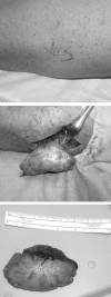
Lipomas are slow-growing soft tissue tumours that rarely reach a size larger than 2 cm. Lesions larger than 5 cm, so-called giant lipomas, can occur anywhere in the body but are seldom found in the upper extremities. The authors present their experiences with eight patients having giant lipomas of the upper extremity. In addition, a review of the literature, and a discussion of the appropriate evaluation and management are included.
Les lipomes sont des tumeurs des tissus mous à croissance lente qui atteignent rarement plus de deux centimètres. Des lésions de plus de cinq centimètres, qu’on appelle lipomes géants, peuvent se former n’importe où sur le corps, mais on les observe rarement sur les membres supérieurs. Les auteurs présentent leur expérience auprès de huit patients ayant un lipome géant d’un membre supérieur. L’article inclut une analyse bibliographique et un exposé de l’évaluation et de la prise en charge pertinentes.
Keywords: Giant lipoma; Liposarcoma; Upper extremity.
Figures


References
-
- Cribb GL, Cool WP, Ford DJ, Mangham DC. Giant lipomatous tumours of the hand and forearm. J Hand Surg [Br] 2005;30:509–12. - PubMed
-
- Phalen GS, Kendrick JI, Rodriguez JM. Lipomas of the upper extremity. A series of fifteen tumors in the hand and wrist and six tumors causing nerve compression. Am J Surg. 1971;121:298–306. - PubMed
-
- Salam GA. Lipoma excision. Am Fam Physician. 2002;65:901–4. - PubMed
-
- Terzioglu A, Tuncali D, Yuksel A, Bingul F, Aslan G. Giant lipomas: A series of 12 consecutive cases and a giant liposarcoma of the thigh. Dermatol Surg. 2004;30:463–7. - PubMed
-
- Celik C, Karakousis CP, Moore R, Holyoke ED. Liposarcomas: Prognosis and management. J Surg Oncol. 1980;14:245–9. - PubMed
LinkOut - more resources
Full Text Sources
