

Frequency Doubling Technology vs Standard Automated Perimetry in Ocular Hypertensive Patients
- PMID: 19554222
- PMCID: PMC2701324
- DOI: 10.2174/1874364100903010006
Frequency Doubling Technology vs Standard Automated Perimetry in Ocular Hypertensive Patients
Abstract
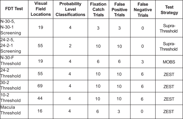
Background: Frequency doubling technology (FDT) perimetry measures contrast sensitivity. The magnocellular component of ganglion cells in human retina is isolated as a whole by the FDT stimulus. The aim of this study is to investigate the role of Humphrey Matrix threshold testing in the detection of early functional retinal impairment in ocular hypertensive patients compared to standard automated perimetry (SAP).
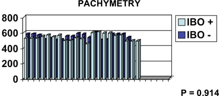
Methodology: Forty hypertensive patients were enrolled in this longitudinal observational clinical study. Functional testing included randomly Humphrey Matrix perimetry and white-on-white Humphrey visual field perimetry. Ibopamine test was performed in all forty patients. The cut-off of 3 mmHg was considered positive for this provocative test.
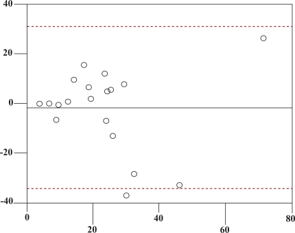
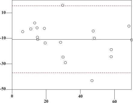
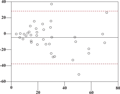
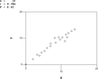
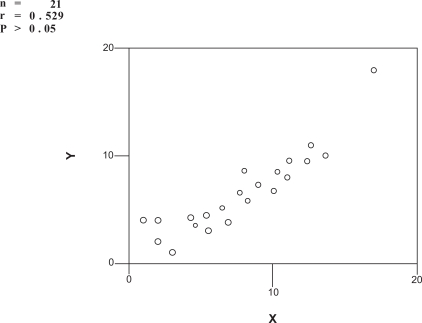
Results: Out of 40 patients, we included 21 in ibopamine positive group and 19 in ibopamine negative group. These two groups are sex- and age-matched. In ibopamine positive group the mean increase in IOP is 4.6 mmHg (ranging from 3 to 10 mmHg). Statistics showed that correlation between FDT and SAP was statistically significant in ibopamine negative group and not statistically significant in ibopamine positive group. Only one patient, coming from IBO + group, converted from ocular hypertension to glaucoma. All the other subjects remained stable in both groups without any therapy and visual field abnormalities.
Conclusions: FDT showed to be more sensitive and specific than SAP mostly in detection of early visual field impairment in ocular hypertensive patients.
Keywords: Early glaucoma; FDT; SAP.; ibopamine; ocular hypertension.
Figures
References
-
- Parravano M, Oddone F, Mineo D, et al. The role of humphrey matrix testing in the early diagnosis of retinopathy in type 1 diabetes. Br J Ophthalmol. 2008;92:1656–60. - PubMed
-
- White AJR, Sun H, Swanson WH, Lee BB. An examination of physiological mechanisms underlying the frequency-doubling illusion. Invest Opthalmol Vis Sci. 2002;43:3590–9. - PubMed
-
- Centofanti M, Fogagnolo P, Oddone F, et al. Learning effect of Humphrey matrix frequency doubling technology perimetry in patients with ocular hypertension. J Glaucoma. 2008;17(6):436–41. - PubMed
-
- Gordon MO, Kass MA. The ocular hypertension treatment study: design and baseline description of the participants. Arch Ophthalmol. 1999;117 (5):573–83. - PubMed
LinkOut - more resources
Full Text Sources
Miscellaneous