

Femoral bone mineral density reflects histologically determined cortical bone volume in hemodialysis patients
- PMID: 19554246
- PMCID: PMC4501027
- DOI: 10.1007/s00198-009-0988-9
Femoral bone mineral density reflects histologically determined cortical bone volume in hemodialysis patients
Abstract
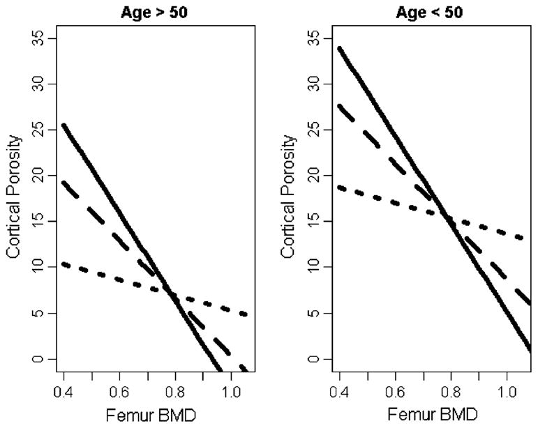
We evaluated the associations between dual energy X-ray absorptiometry (DXA) and histologically determined cancellous and cortical bone volume by controlling for vascular calcifications and demographic variables in hemodialysis (HD) patients. Femoral bone mineral density (f-BMD) was associated with cortical porosity.
Introduction: Assessment of bone mass in chronic kidney disease patients is of clinical importance because of the association between low bone volume, fractures, and vascular calcifications. DXA is used for noninvasive assessment of bone mass whereby vertebral results reflect mainly cancellous bone and femoral results reflect mainly cortical bone. Bone histology allows direct measurements of cancellous and cortical bone volume. The present study evaluates the association between DXA and histologically determined cancellous and cortical bone volumes in HD patients.
Methods: In 38 HD patients, DXA was performed for assessment of bone mass, anterior iliac crest bone biopsies for bone volume, and multislice computed tomography for vascular calcifications.
Results: While lumbar bone mineral density (l-BMD) by DXA was not associated with histologically measured cancellous bone volume, coronary Agatson score showed a borderline statistically significant association (P = 0.055). When controlled for age and dialysis duration, f-BMD by DXA was associated with cortical porosity determined by histology (P = 0.005).
Conclusions: The usefulness of l-BMD for predicting bone volume is limited most probably because of interference by soft tissue calcifications. In contrast, f-BMD shows significant association with cortical porosity.
Conflict of interest statement
Figures
References
-
- Alem AM, Sherrard DJ, Gillen DL, Weiss NS, Beresford SA, Heckbert SR, Wong C, Stehman-Breen C. Increased risk of hip fracture among patients with end-stage renal disease. Kidney Int. 2000;58:396–399. - PubMed
-
- Jadoul M, Albert JM, Akiba T, Akizawa T, Arab L, Bragg-Gresham JL, Mason N, Prutz KG, Young EW, Pisoni RL. Incidence and risk factors for hip or other bone fractures among hemodialysis patients in the dialysis outcomes and practice patterns study. Kidney Int. 2006;70:1358–1366. - PubMed
-
- Schulz E, Arfai K, Liu X, Sayre J, Gilsanz V. Aortic calcification and the risk of osteoporosis and fractures. J Clin Endocrinol Metab. 2004;89:4246–4253. - PubMed
-
- Coco M, Rush H. Increased incidence of hip fractures in dialysis patients with low serum parathyroid hormone. Am J Kidney Dis. 2000;36:1115–1121. - PubMed
Publication types
MeSH terms
Grants and funding
LinkOut - more resources
Full Text Sources
Medical
