

Review
doi: 10.1007/s00018-009-0038-y.
Epub 2009 May 12.
Kank proteins: structure, functions and diseases
Affiliations
- PMID: 19554261
- PMCID: PMC11115667
- DOI: 10.1007/s00018-009-0038-y
Item in Clipboard
Review
Kank proteins: structure, functions and diseases
Cell Mol Life Sci.
2009 Aug.
Abstract
The Kank family of proteins, Kank1-Kank4, are characterized by their unique structure, coiled-coil motifs in the N-terminal region, and ankyrin-repeats in the C-terminal region, with an additional motif, the KN motif, at the N-terminus. Kank1 was obtained by positional cloning of a tumor suppressor gene in renal cell carcinoma, while the other members were found by homology search. The family is involved in the regulation of actin polymerization and cell motility through signaling pathways containing PI3K/Akt and/or unidentified modulators/effectors. Their relationship to diseases such as cancer, and to neuronal and developmental disorders, will be an important subject of future study.
Figures

Structure and functions of human Kank proteins. a Schematic structure of human Kank proteins. The human Kank family (KANK1, KANK2, KANK3, and KANK4) share a common structural feature; the Kank N-terminal (KN) motif (orange boxes), and coiled-coil and ankyrin-repeat domains from the N- to C-terminal. The coiled-coil domain contains coiled-coil motifs (green boxes) and the ankyrin-repeat domain contains ankyrin-repeats (black boxes). The variants of the coiled-coil motifs are numbered. b Inhibition of the formation of actin stress fibers by overexpression of Kank proteins in NIH3T3 cells. Each of the Kank proteins was detected by immunostaining (green). Actin stress fibers were detected with rhodamine-conjugated phalloidin (red). Cells expressing FLAG-GST were used as a control. The cells expressing Kank proteins or FLAG-GST are indicated by arrowheads. Scale bars 20 μm
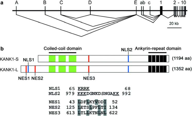
Structure of the Kank1 gene and Kank1 protein. a The genomic organization of the human Kank1 gene (KANK1). The alternative exons detected in the normal human kidney are labeled in upper case letters, whereas the alternative exons detected in VMRC-RCW cells are labeled in lower case. b Schematic structure of the human KANK1-L and KANK1-S proteins (KANK1-L and KANK1-S), showing identified domains and motifs. The sequences of NLS (NLS1 and NLS2) and NES (NES1 to NES3) motifs identified in KANK1-L are shown, with conserved sites underlined (NLSs) or shaded (NESs)

Inhibition of cell migration by Kank1. Time-lapse confocal laser microscopic images of the tetracycline-inducible NIH3T3 cells are shown in the six left panels. The expression of GFP-Kank1 was induced with doxycycline (+DOX; lower panels), while the control cells were treated with ethanol (−DOX; upper panels). The images were taken every 20 min. The images are fluorescence micrographs merged with phase-contrast micrographs, with the cells containing GFP-Kank1 shown in green. The migration of cells was traced (right-most panels), where the centers of the nuclei are marked. Solid arrows indicate the direction of cell movement. Scale bars 10 μm
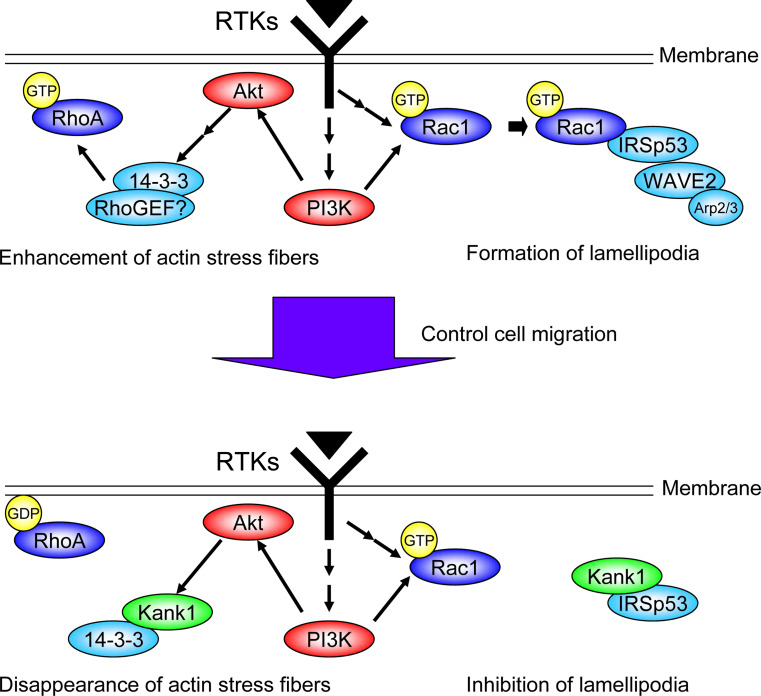
A model of the role of Kank1. A hypothetical model of the function of Kank1 at the leading edge of cells is shown. When cells are moving, stimulated RTKs activate Rac1, and active Rac1 (GTP-loaded form of Rac1) associates with the IRSp53/WAVE2/Arp2/3 complex, resulting in the formation of lamellipodia (upper, right). Stimulated RTKs also activate RhoA through a complex containing 14-3-3, and active RhoA (GTP-loaded form of RhoA) enhances the development of actin stress fibers in the leading edge (upper, left). When the cells need to control the migration or stop the movement, Kank1 is activated at the leading edge or translocated there, where it associates with IRSp53 and competes with active Rac1 to bind to IRSp53 (lower, right). Kank1 is also phosphorylated by Akt during this process, secludes 14-3-3 from an activation complex for RhoA, resulting in the inhibition of RhoA, and thereby decreases the formation of actin stress fibers at the leading edge (lower, left). Both these signal pathways could be inhibited by Kank1
References
-
- Zhu Y, Kakinuma N, Wang Y, Kiyama R. Kank proteins: a new family of ankyrin-repeat domain-containing proteins. Biochim Biophys Acta. 2008;1780:128–133. - PubMed
-
- Rodley P, Hatano N, Nishikawa NS, Roy BC, Sarkar S, Kiyama R. A differential genomic cloning method for cancer study: an outline and applications. Recent Res Dev Mol Biol. 2003;1:13–27.
Publication types
MeSH terms
Substances
LinkOut - more resources
Full Text Sources
