. 2010 May;20(5):596-607.
doi: 10.1002/hipo.20665.

Alcohol inhibition of neurogenesis: a mechanism of hippocampal neurodegeneration in an adolescent alcohol abuse model

Affiliations

Alcohol inhibition of neurogenesis: a mechanism of hippocampal neurodegeneration in an adolescent alcohol abuse model

Stephanie A Morris et al. Hippocampus. 2010 May.

Abstract

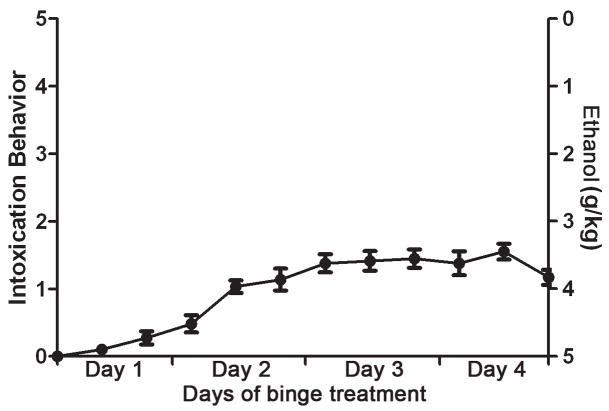
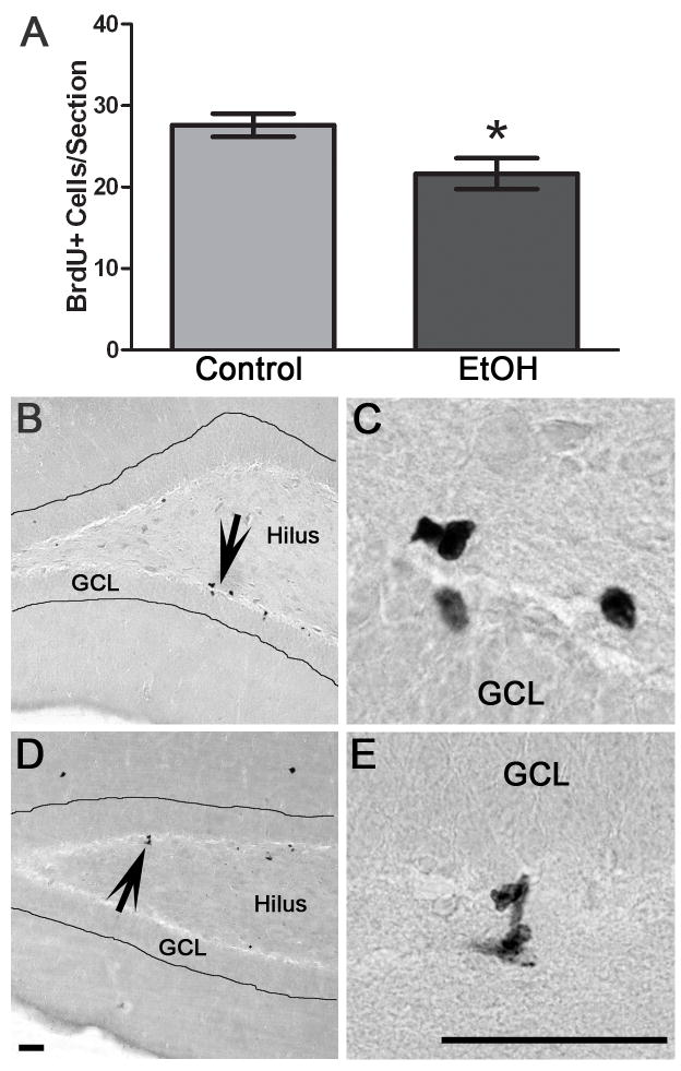
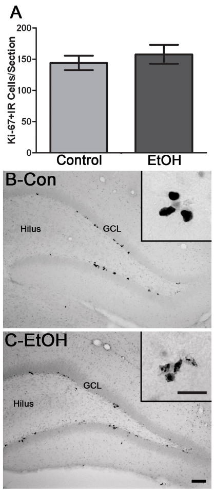
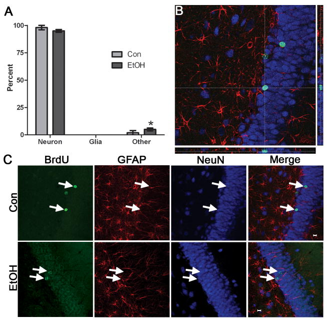
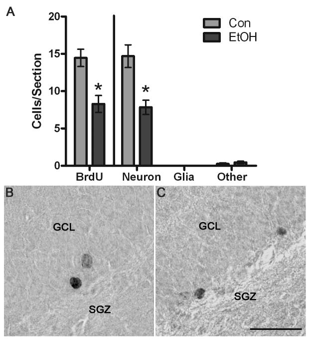
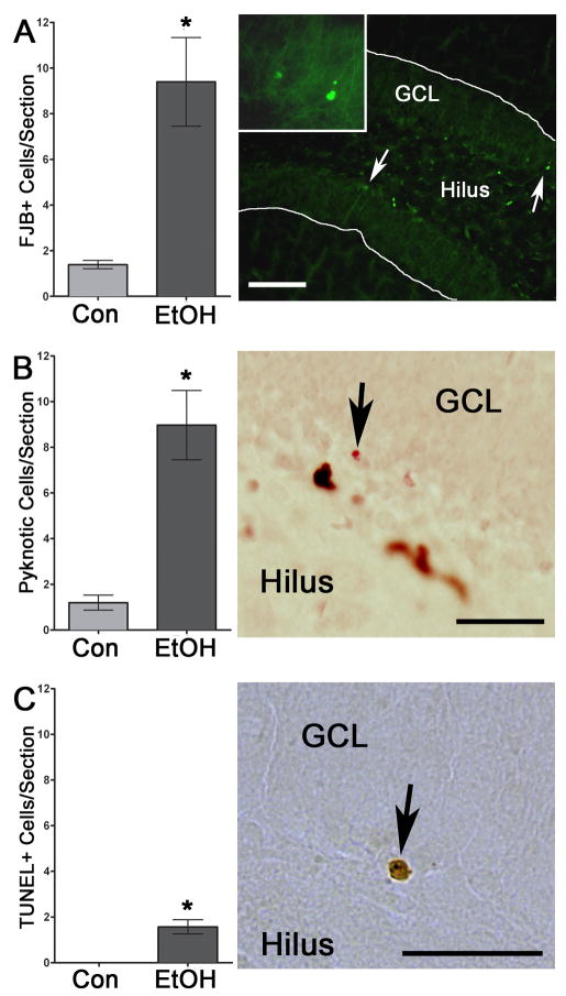
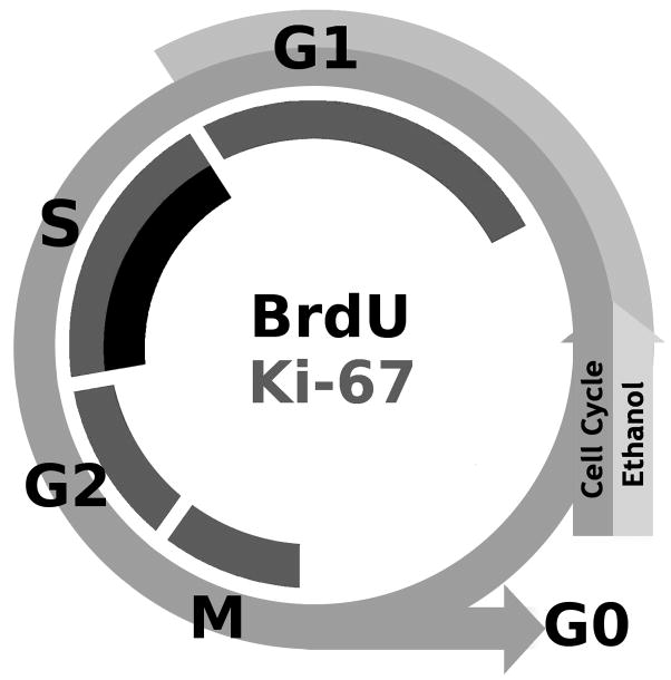
Adolescents diagnosed with an alcohol use disorder show neurodegeneration in the hippocampus, a region important for learning, memory, and mood regulation. This study examines a potential mechanism by which excessive alcohol intake, characteristic of an alcohol use disorder, produces neurodegeneration. As hippocampal neural stem cells underlie ongoing neurogenesis, a phenomenon that contributes to hippocampal structure and function, we investigated aspects of cell death and cell birth in an adolescent rat model of an alcohol use disorder. Immunohistochemistry of various markers along with Bromo-deoxy-Uridine (BrdU) injections were used to examine different aspects of neurogenesis. After 4 days of binge alcohol exposure, neurogenesis was decreased by 33 and 28% at 0 and 2 days after the last dose according to doublecortin expression. To determine whether this decrease in neurogenesis was due to effects on neural stem cell proliferation, quantification of BrdU-labeled cells revealed a 21% decrease in the dentate gyrus of alcohol-exposed brains. Cell survival and phenotype of BrdU-labeled cells were assessed 28 days after alcohol exposure and revealed a significant, 50% decrease in the number of surviving cells in the alcohol-exposed group. Reduced survival was supported by significant increases in the number of pyknotic-, FluoroJade B positive-, and TUNEL-positive cells. However, so few cells were TUNEL-positive that cell death is likely necrotic in this model. Although alcohol decreased the number of newborn cells, it did not affect the percentage of cells that matured into neurons (differentiation). Thus, our data support that in a model of an adolescent alcohol use disorder, neurogenesis is impaired by two mechanisms: alcohol-inhibition of neural stem cell proliferation and alcohol effects on new cell survival. Remarkably, alcohol inhibition of neurogenesis may outweigh the few dying cells per section, which implies that alcohol inhibition of neurogenesis contributes to hippocampal neurodegeneration in alcohol use disorders.


Figures

Figure 1
Figure 1
Alcohol (ethanol) is administered in a four day binge model as reported previously (Nixon & Crews, 2004). At each feeding (7am, 3pm, 11pm), behavioral intoxication is scored using a scale (0–5) modified from Majchrowicz (1975) and a corresponding dose of ethanol (0g/kg–5g/kg) administered by gavage. The mean intoxication score (left axis) for all ethanol-exposed rats is plotted and shows that adolescent rats only show mild ataxia. Values shown are mean ± SEM.
Figure 2
Figure 2
Binge alcohol administration reduces DCX+IR during and following alcohol intoxication in adolescent rats. A) DCX+IR is significantly decreased following four days of binge exposure to alcohol (4D; Con n=6, EtOH n=6) and two days after the last dose of alcohol (4D+2; Con n=7, EtOH n=8). DCX+IR returns to control levels by 7 days after the last dose (4D+7; Con n=7, EtOH n=8). DCX is expressed between several hours to a few days after a cell divides such that its expression reflects activities that alter neurogenesis days prior (Brown et al., 2003). Representative photomicrographs of DCX expression in the dentate gyrus are shown for control (Con, B) and alcohol (EtOH, C) exposure at the 4D timepoint. Scale bar = 100μm. Inset scale bar = 20μm *p< 0.05
Figure 3
Figure 3
Binge alcohol administration reduces the number of BrdU+ cells, or cell proliferation, during alcohol intoxication in adolescent rats. A) To label cell proliferation, BrdU was injected 1 h after the last dose of alcohol or control diet and rats were sacrificed 2 h later. The number of BrdU+ cells is significantly decreased in binge alcohol exposed rats (EtOH, n=4) versus controls (n=5). Representative photomicrographs of BrdU+ clusters scattered along both blades of the dentate gyrus of the hippocampus are shown for control (B) and alcohol rats (D). Higher magnification images show representative BrdU+ cells from control (C) and alcohol-treated rats (E). GCL= Granule cell layer. Scale bars = 50 μm. *p< 0.05
Figure 4
Figure 4
Binge alcohol administration does not change the number of Ki-67+ cells during alcohol intoxication in adolescent rats. A) The number of Ki-67+ cells is identical between binge alcohol exposed rats (EtOH, n=6) and controls (n=6) which supports that binge alcohol exposure does not alter the number of cells in the active phases of the cell cycle. Representative photomicrographs show that Ki-67+ clusters are scattered along both blades of the dentate gyrus of the hippocampus in control (B) and alcohol-treated rats (C). GCL= Granule cell layer. Scale bars = 100μm. Inset scale bar = 30μm. *p< 0.05
Figure 5
Figure 5
Binge alcohol administration does not alter the phenotype of cells born during alcohol intoxication. BrdU was injected at 4D and rats were sacrificed 28 days later (4D+28). A) Triple fluorescent labeling for BrdU, a neuron-specific marker (NeuN) and an astroglia specific marker (GFAP) was assessed for colabeling in 50 BrdU+ cells per subject. The percentage of BrdU+ cells that expressed a neuronal phenotype (BrdU+/NeuN+) or an astroglial phenotype (BrdU+/GFAP+) was similar in control (n=5) and alcohol-exposed tissue (n=4). There was only a significant increase in “other” cells (BrdU+/NeuN-/GFAP-) in the alcohol-exposed tissue, which merely shows that other types of cells are proliferating and remaining after binge treatment. These few other cells could be microglia, endothelial cells, or oligodendroglia. A representative orthogonal view of reconstructed Z-stacks is shown in B where BrdU is labeled in green, NeuN in blue, and GFAP in red. The crosshairs are placed atop the cell of interest such that colabeling can be assessed from all planes. C. Representative images of individual fluorochromes showing BrdU+ cells (green), GFAP+ cells (red), NeuN+ cells (blue), and merged in both the control and alcohol-exposed tissue.
Figure 6
Figure 6
BrdU+ cells remain significantly decreased 28 d after the last dose of alcohol. A) In the dorsal dentate gyrus granule cell layer and subgranular zone, there were significantly fewer BrdU+ cells in the alcohol-exposed tissue (n=4), as compared to controls (n=5) as shown on the left side of the graph. Because BrdU alone does not indicate cell type, BrdU+ cell numbers were multiple multiplied by the phenotype percentage data (Figure 5) to estimate effects on neurogenesis, which is shown in the right portion of the graph. Thus, the significant decrease in BrdU+ cells, yet similar phenotypes, result in significantly fewer neurons. Representative photomicrographs of BrdU+ cells labeled with DAB are shown for control tissue (B) and in alcohol-exposed tissue (C). Scale bar = 30 μm. *p< 0.05
Figure 7
Figure 7
Cell death is significantly increased after four days of binge alcohol exposure according to three different measures. A) Binge alcohol exposure significant increased the number of FJB+ cells in the dentate gyrus as shown in representative photomicrographs of the alcohol-exposed tissue. Inset picture is of cells noted by the right most arrow. Scale bar = 100μm. B) Similarly, pyknosis was significantly increased after alcohol exposure in the dentate gyrus and subgranular zone. Arrows indicate a cell with condensed chromatin in the nucleus, characteristic of a pyknosis from a representative Ki67-labeled section (brown cells) that was counterstained in Neutral Red (pink). Scale bar = 30μm. C) The mechanism of cell death was examined by TUNEL staining for apoptosis. Very little TUNEL reactivity was observed, even though it was slightly but significantly increased after binge alcohol treatment, which suggests that cell death observed by FJB and pyknosis is not apoptotic. Past work in binge-exposed adults supports this observation (Obernier et al., 2002) as adult rats were similarly TUNEL negative and morphological assessments indicated dark cell degeneration, a necrotic form of cell death. Arrow indicates one of the very few TUNEL+ cells in the subgranular zone of the dentate gyrus. Scale bar = 30 μm. *p< 0.05
Figure 8
Figure 8
A representation of the two markers used to assess proliferating cells in comparison with the stages of the cell cycle. Ki-67, an endogenous marker of proliferation is expressed during all active phases of the cell cycle (M, G2, S, and G1; Scholzen and Gerdes, 2000). BrdU, an exogenous marker, is incorporated into cells when the DNA is single stranded, which occurs during the S-phase of the cell cycle. Although cells arrested in G1 or G2 phases are considered mitotically active and express Ki67, they would not incorporate BrdU. The outer arrow depicts the cell cycle in ethanol-exposed cells arrested in G1 phase, where cells would express Ki-67 but not incorporate BrdU.

Similar articles

Cited by

References

    1. Altman J, Das GD. Autoradiographic and histological evidence of postnatal hippocampal neurogenesis in rats. J Comp Neurol. 1965;124(3):319–35. - PubMed
    1. Bechara A. Decision making, impulse control and loss of willpower to resist drugs: a neurocognitive perspective. Nat Neurosci. 2005;8(11):1458–63. - PubMed
    1. Brown JP, Couillard-Despres S, Cooper-Kuhn CM, Winkler J, Aigner L, Kuhn HG. Transient expression of doublecortin during adult neurogenesis. J Comp Neurol. 2003;467(1):1–10. - PubMed
    1. Cameron HA, McKay RD. Adult neurogenesis produces a large pool of new granule cells in the dentate gyrus. J Comp Neurol. 2001;435(4):406–17. - PubMed
    1. Canales JJ. Adult neurogenesis and the memories of drug addiction. Eur Arch Psychiatry Clin Neurosci 2007 - PubMed

Publication types

MeSH terms