

Nanoscale growth factor patterns by immobilization on a heparin-mimicking polymer
- PMID: 19554729
- PMCID: PMC3110987
- DOI: 10.1021/ja803676r
Nanoscale growth factor patterns by immobilization on a heparin-mimicking polymer
Abstract
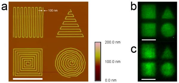
In this study, electrostatic interactions between sulfonate groups of an immobilized polymer and the heparin binding domains of growth factors important in cell signaling were exploited to nanopattern the proteins. Poly(sodium 4-styrenesulfonate-co-poly(ethylene glycol) methacrylate) (pSS-co-pPEGMA) was synthesized by reversible addition-fragmentation chain transfer (RAFT) polymerization using ethyl S-thiobenzoyl-2-thiopropionate as a chain transfer agent and 2,2'-azoisobutyronitrile (AIBN) as the initiator. The resulting polymer (1) was characterized by 1H NMR, GPC, FT-IR, and UV-vis and had a number average molecular weight (Mn) of 24,000 and a polydispersity index (PDI) of 1.17. The dithioester end group of 1 was reduced to the thiol, and the polymer was subsequently immobilized on a gold substrate. Binding of basic fibroblast growth factor (bFGF) and vascular endothelial growth factor (VEGF) to the polymer via the heparin binding domains was then confirmed by surface plasmon resonance (SPR). The interactions were stable at physiological salt concentrations. Polymer 1 was cross-linked onto silicon wafers using an electron beam writer forming micro- and nanopatterns. Resolutions of 100 nm and arbitrary nanoscale features such as concentric circles and contiguous squares and triangles were achieved. Fluorescence microscopy confirmed that bFGF and VEGF were subsequently immobilized to the polymer micro- and nanopatterns.
Figures


References
-
- Jung DR, Kapur R, Adams T, Giuliano KA, Mrksich M, Craighead HG, Taylor DL. Crit. Rev. Biotechnol. 2001;21:111–154. - PubMed
-
- Refai AK, Textor M, Brunette DM, Waterfield JD. J. Biomed. Mater. Res. A. 2004;70A:194–205. - PubMed
-
- Falconnet D, Csucs G, Grandin HM, Textor M. Biomaterials. 2006;27:3044–3063. - PubMed
-
- Arnold M, Cavalcanti-Adam EA, Glass R, Blummel J, Eck W, Kantlehner M, Kessler H, Spatz JP. Chemphyschem. 2004;5:383–388. - PubMed
-
- Lee KB, Park SJ, Mirkin CA, Smith JC, Mrksich M. Science. 2002;295:1702–1705. - PubMed
Publication types
MeSH terms
Substances
Grants and funding
LinkOut - more resources
Full Text Sources
Other Literature Sources
Medical
