. 2008 Dec 10;130(49):16778-85.
doi: 10.1021/ja805570f.

Functionalized single-walled carbon nanotubes as rationally designed vehicles for tumor-targeted drug delivery

Affiliations

Functionalized single-walled carbon nanotubes as rationally designed vehicles for tumor-targeted drug delivery

Jingyi Chen et al. J Am Chem Soc. .

Abstract

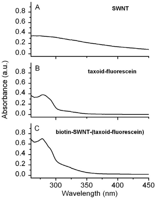
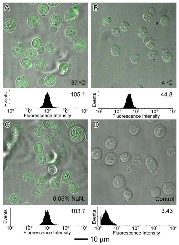
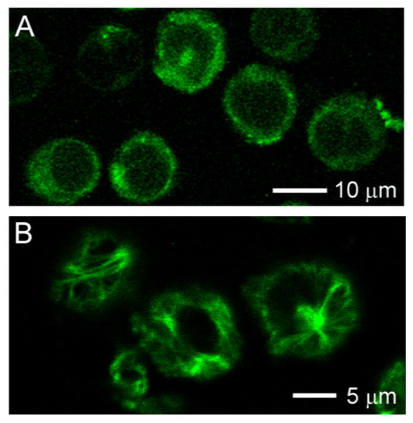
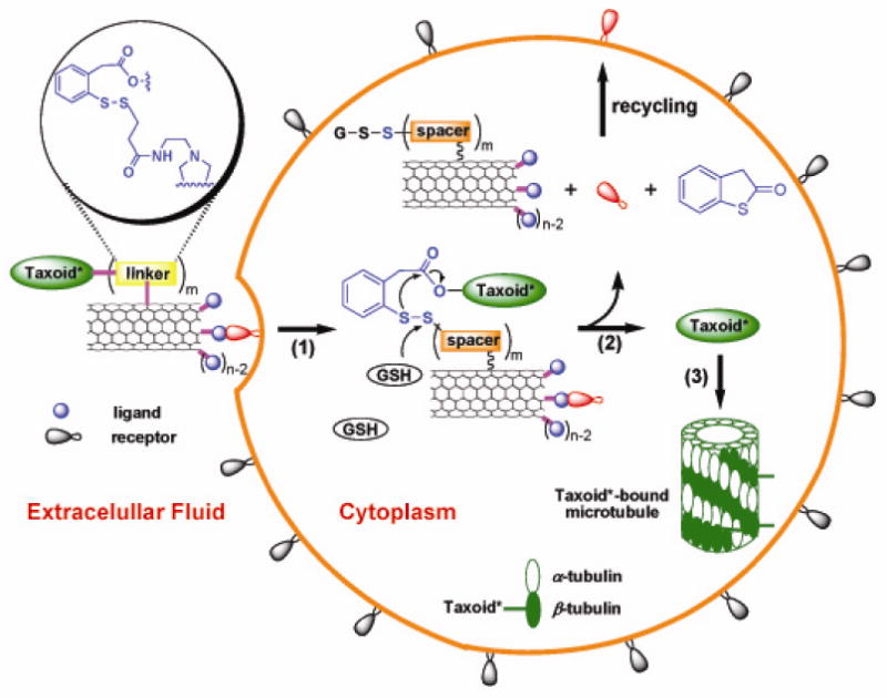
A novel single-walled carbon nanotube (SWNT)-based tumor-targeted drug delivery system (DDS) has been developed, which consists of a functionalized SWNT linked to tumor-targeting modules as well as prodrug modules. There are three key features of this nanoscale DDS: (a) use of functionalized SWNTs as a biocompatible platform for the delivery of therapeutic drugs or diagnostics, (b) conjugation of prodrug modules of an anticancer agent (taxoid with a cleavable linker) that is activated to its cytotoxic form inside the tumor cells upon internalization and in situ drug release, and (c) attachment of tumor-recognition modules (biotin and a spacer) to the nanotube surface. To prove the efficacy of this DDS, three fluorescent and fluorogenic molecular probes were designed, synthesized, characterized, and subjected to the analysis of the receptor-mediated endocytosis and drug release inside the cancer cells (L1210FR leukemia cell line) by means of confocal fluorescence microscopy. The specificity and cytotoxicity of the conjugate have also been assessed and compared with L1210 and human noncancerous cell lines. Then, it has unambiguously been proven that this tumor-targeting DDS works exactly as designed and shows high potency toward specific cancer cell lines, thereby forming a solid foundation for further development.


Figures

Figure 1
Figure 1
TEM images of HiPco SWNTs: (A) pristine SWNTs; (B) acid oxidized SWNTs. (C) AFM height image of acid-oxidized SWNTs 4, and (D) ATR-IR spectra of SWNTs vs. acid oxidized SWNTs. Note that the peak at 2349 cm-1 can be attributed to the asymmetric stretching mode of CO2 molecules in the atmosphere.
Figure 2
Figure 2
UV-visible spectra of SWNTs and their biofunctionalized conjugates plotted to scale: (A) acid oxidized SWNTs 4 at 50 μg/mL concentration; (B) taxoid-fluorescein conjugates at 15 μM concentration; and (C) biotin-SWNT-(taxoid-fluorescein) conjugate 3 at 50 μg/mL concentration.
Figure 3
Figure 3
(A) Photographs of vials containing pristine SWNT 0, SWNT-FITC 1, biotin-SWNT-FITC 2, and biotin-SWNT-(taxoid-fluorescein) 3 in CH2Cl2 (∼1 mg/mL). (B) Photographs of SWNT conjugates at concentrations of 50 μg/mL in cell culture medium: SWNT-FITC conjugate 1 (left) and biotin-SWNT-(taxoid-fluorescein) conjugate 3 (right). Arrow indicates that some of the nanotubes 1 precipitated from the medium.
Figure 4
Figure 4
CFM images of L1210FR cells after incubation with SWNT-FITC 1 (A) and biotin-SWNT-FITC 2 (B) at the final concentration of 10 μg/mL at 37 °C for 2 h. (C) Comparison of fluorescence intensities of L1210FR cells by flow cytometry upon treatment with pristine SWNTs 0 (purple), conjugate 1 (blue), and conjugate 2 (red) at the final concentration of 10 μg/mL in each case. Background, i.e., data for untreated cells, is plotted in black.
Figure 5
Figure 5
CFM images and flow cytometry analysis of L1210FR cells after incubation with SWNT-FITC 1 at the final concentration of 10 μg/mL under different conditions after 3 h of incubation: (A) at 37°C; (B) at 4 °C; and (C) at 37 °C in the presence of 0.05% (w/v) NaN3. (D) Control experiment: CFM images and flow cytometry data of L1210FR cells after treatment with oxidized SWNT 4 at the same concentrations at 37 °C for 3 h. All of the CFM images and flow cytometry data were acquired under the same conditions.
Figure 6
Figure 6
CFM images and flow cytometry analysis of L1210FR cells after incubation with biotin-SWNT-FITC 2 at a final concentration of 10 μg/mL under different conditions after an incubation period of 3 h: (A) at 37°C; (B) at 4 °C; (C) at 37 °C in the presence of 0.05% (w/v) NaN3; (D) at 37 °C after pretreatment with excess biotin. All of the CFM images and flow cytometry data were acquired under the same conditions.
Figure 7
Figure 7
CFM images of L1210FR cells treated with biotin-SWNT-(taxoid-fluorescein) 3 incubated before (A) and after (B) addition of GSH-ethyl ester. Image (B) clearly highlights the presence of fluorescent microtubule networks in the living cells generated by the binding of the fluorescent taxoid (SB-T-1214-fluorescein) upon cleavage of the disulfide bond in the linker by either GSH or GSH-ethyl ester.
Figure 8
Figure 8
CFM images and flow cytometry analysis of different cell types upon incubation with biotin-SWNT-taxoid conjugate 3 at a final concentration of 50 μg/mL at 37 °C for an incubation period of 3 h: (A) L1210FR leukemia cell line overexpressing biotin receptors; (B) L1210 leukemia cell line; and (C) WI38 human lung fibroblast cell line. All of the CFM images and flow cytometry data were taken under identical conditions.
Scheme 1
Scheme 1
Schematic illustration of three key steps involved in the tumor-targeting drug delivery of biotin-SWNT-taxoid conjugate 3: (1) internalization of the whole conjugate via receptor-mediated endocytosis; (2) drug release through cleavage of the disulfide linker moiety by intracellular thiol, e.g., GSH; (3) binding of the free taxoid molecules to tubulins/microtubules, forming stabilized microtubules that block cell mitosis and trigger apoptosis. [Note: Since each taxoid molecule is fluorescently labeled with fluorescein, the internalized conjugates 3 in the cytoplasm and the taxoid-bound microtubules are fluorescent.]
Scheme 2
Scheme 2
Synthesis of SWNT-FITC conjugate 1.
Scheme 3
Scheme 3
Synthesis of biotin-SWNT-FITC conjugate 2.
Scheme 4
Scheme 4
Synthesis of biotin-SWNT-linker-(taxoid-fluorescein) conjugate 3.

References

    1. Leuschner C, Kumar C. In: Nanofabrication Towards Biomedical Application. Kumar CSSR, Jormes J, Leuschner C, editors. Wiley-VCH; 2005. pp. 289–326.
    1. Ferrari M. Nature Rev. 2005;5:161–171. - PubMed
    1. Lacerda L, Bianco A, Prato M, Kostarelos K. Adv Drug Delivery Rev. 2006;58:1460–1470. - PubMed
    1. Dresselhaus M, Dai H. MRS 2004 Carbon Nanotube Special Issue. 2004.
    1. Dai H. Surf Sci. 2002;500:218–241.

Publication types